The small intestine is a coiled tube that extends from the pylorus to the ileocaecal valve, where it continues into the large intestine.
Its average length in an adult is about 5 metres.
It lies in the central and lower part of the abdominal cavity, generally within the boundaries marked out by the large intestine (the colic frame).
The small intestine has two parts:
- The duodenum: a short, fixed part with no mesentery, which is closely related to the pancreas, forming the duodenopancreatic complex (already covered).
- The jejunum and ileum: a long, coiled, mobile part that is attached to the posterior wall of the abdomen by the mesentery.

Remember that this lesson is part of the free courses from Paradigmia. Access the full course below 👇👇 HUMAN ANATOMY COURSE
Jejunum and ileum
Together they form the longest part of the small intestine (the tenue), where most food absorption takes place. They also produce hormones that regulate intestinal secretion and motility, and their walls contain numerous lymphoid formations with an immune function.
This part of the small intestine extends from the duodenojejunal flexure to the ileocaecal junction.
The jejunum and ileum are characterised mainly by:
- Their length, so that in the abdominal cavity they form numerous windings, giving rise to the intestinal loops.
- Their mobility, made possible by the length of the mesentery, which attaches them to the posterior wall of the abdomen.
- The importance of their digestive functions, which is why they have a rich arterial and venous blood supply and extensive lymphatic drainage.
Externally there is nothing to tell them apart except their position. The jejunum lies in the upper part, near the duodenojejunal angle, whereas the ileum lies lower down and to the right, near the ileocaecal flexure.
Unrolled, it appears as a tube of variable length, whose calibre decreases from its origin (4 cm) to its end (2-3 cm).
One variation seen in 2% of the population is a finger-of-a-glove-shaped outpouching about 80 cm from the ileocaecal angle. This outpouching is called the ileal (or Meckel’s) diverticulum and represents an embryological remnant of the omphalomesenteric (vitelline) duct (the duct that joined the primitive gut to the umbilicus).

The jejunum and ileum are made up of the three layers common to the intra-abdominal digestive system:
The mucosa
It is of the intestinal type: the inner surface of the intestine has numerous projections called circular folds, which project into the lumen, are permanent and do not disappear when the intestine is full.

In addition, its surface has a velvety appearance because of tiny microscopic projections, the intestinal villi and microvilli. Between the villi there are millions of openings into which the intestinal glands drain, secreting mucus and intestinal juice.

The intestinal mucosa is also an immune organ and contains lymphoid formations, which can be seen either singly (solitary follicles) or grouped into patches (Peyer’s patches). The features of the intestine change progressively. The circular folds are abundant in the duodenum and jejunum (proximal half) and from there decrease considerably, almost disappearing completely in the distal ileum. Their function is to slow the passage of food and increase the absorptive surface.
The muscular layer
It is smooth muscle forming two layers: a superficial layer of longitudinal fibres and a deeper layer of circular fibres. It is responsible for the mixing (or segmentation) movements and the peristaltic (propulsive) movements.
The peritoneum
They are intraperitoneal organs: the visceral peritoneum surrounds the jejunum and ileum and then continues as the mesentery, which fixes them to the posterior wall.
In addition, between the layers of the intestinal wall lie the intramural nerve plexuses, which regulate the motility of the intestine (they will be covered with the innervation of the intestine).
The mesentery
The mesentery is a fan-shaped peritoneal fold that attaches the jejunum and ileum to the posterior wall of the abdomen and consists of:
- Two layers or surfaces.
- Two borders: visceral (intestinal attachment) and parietal (parietal attachment, or root).
- Contents: superior mesenteric vessels, nerves, lymph nodes and fat.
Let’s look at this image from Mayo Clinic , where it is well explained:
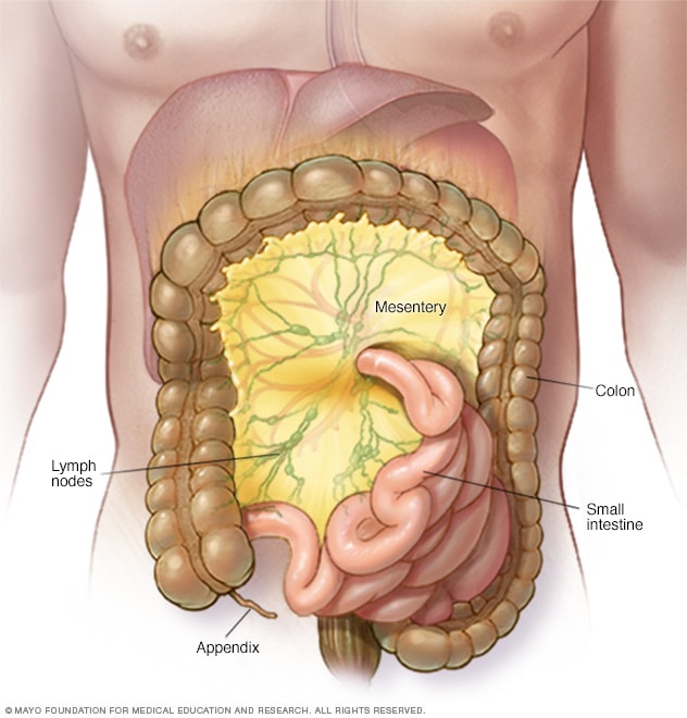
The root of the mesentery is the border that attaches it to the parietal peritoneum of the posterior wall of the abdomen. It measures about 15 cm and runs obliquely, from the duodenojejunal junction to the ileocaecal junction. On its way, the root of the mesentery crosses in front of the following retroperitoneal structures in turn:
- The duodenum: 3rd and 4th parts.
- Abdominal aorta and inferior vena cava.
- Right ureter, right ovarian or testicular vessels and iliopsoas muscle.
Relations of the small intestine
The jejunum and ileum lie in the abdominopelvic cavity, in the infracolic space, within the colic frame. Their main relations are:
- Anteriorly: the anterior abdominal wall, through the greater omentum.
- Posteriorly, through the parietal peritoneum, retroperitoneal structures such as the infracolic part of the duodenopancreatic complex, the abdominal aorta, the inferior vena cava and the ureters.
- Inferiorly: the pelvic viscera: the rectum and bladder in men and, in women, also the uterus.
Differences between the jejunum and the ileum
To finish this lesson on the small intestine, we will review some of the differences between the jejunum and the ileum:
- Position and arrangement: The loops of the jejunum are arranged horizontally and lie closer to the duodenojejunal flexure, whereas the loops of the ileum are arranged vertically and lie closer to the ileocaecal flexure.
- Calibre: calibre decreasing from proximal to distal (the jejunum has the larger calibre).
- Mesentery: in the ileum it is thicker because it contains more fat.
- The intestinal wall: in the jejunum it is thicker, with more circular folds and with solitary follicles. In the ileum, Peyer’s patches appear.
To finish this lesson, in the following video we will look at some of the anatomical features of the jejunum and ileum.
Remember that this lesson is part of the free courses from Paradigmia. Access the full course below 👇👇 HUMAN ANATOMY COURSE