It is the last part of the digestive tract, continuing from the ileum. It extends from the ileocaecal junction to the anal orifice. It lies in the abdominal and pelvic cavities.
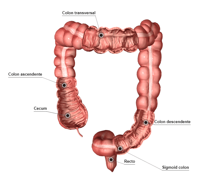
Three segments can be distinguished in the large intestine: the caecum, the colon and the rectum.
- The caecum, to which the appendix is attached, lies below the ileocaecal junction.
- The colon takes up a position around the jejunum and ileum, called the "colic frame", and is divided into four parts:
- The ascending colon: lying on the right side of the abdomen, it runs towards the inferior surface of the liver, where it forms the right (hepatic) colic flexure or angle.
- The transverse colon: which crosses the abdomen as far as its upper left part, where, beneath the spleen, it forms the left (splenic) colic flexure or angle.
- The descending colon: runs vertically down to the left iliac crest.
- The sigmoid colon: is the terminal segment of the colon and extends from the descending colon into the lesser pelvis (at the level of the 3rd sacral vertebra).
- The rectum: descends in front of the sacrum and opens to the exterior at the anus.
External features
The large intestine measures about 1.5 metres. Its calibre decreases from the caecum (7-8 cm) to the sigmoid colon (3-5 cm), while the rectum is initially dilated into an ampulla and then narrows.
It differs from the small intestine, besides being bulkier, in three main features:
Taeniae coli, or longitudinal bands of the colon
These structures are formed by longitudinal muscle fibres, which group together to form cords that follow the long axis of the intestine. The three taeniae originate at the base of the appendix and diverge from there along the colon.
Haustra or sacculations
They are separated by grooves (semilunar folds) that project into the lumen of the colon. The haustra disappear at the level of the rectum.
Omental (epiploic) appendices
These are small, more or less pedunculated masses of fat that hang along the taeniae. They are absent from the caecum and the rectum.
Anatomical structure
Three layers can be seen in the wall of the large intestine:
The mucosa
It lacks the circular folds and villi of the small intestine, but it does have longitudinal and transverse folds, which correspond to the grooves produced by the relief of the taeniae and haustra.
The muscular layer
As in the small intestine, it consists of two layers: an outer layer of longitudinal fibres that group together into the taeniae, and an inner layer of circular fibres, which form the semilunar folds and encircle the circumference of the colon.
The serosa, the peritoneum
Its arrangement varies with each segment of the large intestine.
Caecum and appendix
The caecum is the first part of the large intestine, lying below the ileocaecal junction, in the right iliac fossa. It has an extension, the appendix, which can often become inflamed (appendicitis), giving the caecum and appendix together considerable clinical interest. The caecum acts as a reservoir for the faecal bolus, which is held up and thickened by the reabsorption of water. By means of contractions, the caecum pushes the bolus into the ascending colon.
The caecum is a sac open at the top, measuring about 6 cm in height and 6-8 cm in width, with a rounded bottom below.
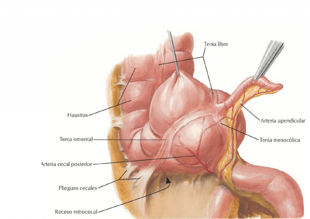
On its inner surface (in the upper part) there are two features: the ileocaecal orifice and, two or three cm below this orifice, the appendix is attached, with the three longitudinal taeniae beginning around it.
Vermiform appendix (vermicular or caecal)
It is tubular, more or less coiled and worm-shaped, and is attached to the medial surface of the caecum, below the ileocaecal orifice. Its length averages about 9 cm, with a diameter of 4-8 mm.
The position of the appendix in relation to the caecum varies: descending, retrocaecal or subcaecal.
The most widely used landmark for marking the depth of the base of the appendix is the junction between the lateral third and the medial two thirds of the line joining the right anterior superior iliac spine to the umbilicus (McBurney’s point).

The function of the appendix is highly debatable. In children it is a lymphatic organ and has important immunological functions. With age, this lymphoid tissue atrophies and is replaced by connective tissue. Inflammation of the appendix (appendicitis) is one of the most frequent causes of acute abdomen and is usually due to obstruction of the appendix by faecal matter.
The pain of acute appendicitis usually begins in the periumbilical region and then settles in the right lower quadrant. In typical appendicitis, pressing with a finger on McBurney’s point causes intense abdominal pain. Appendicitis is treated surgically (appendicectomy), because rupture of the inflamed appendix causes a partial or total infection of the peritoneum (peritonitis), with increased abdominal pain and abdominal rigidity.
Ileocaecal valve
The opening between the ileum and the caecum is bordered by the ileal papilla or ileocaecal valve (of Bauhin), formed by two overlapping lips (upper and lower), which open periodically so that the contents of the small intestine can pass into the large intestine and which also oppose reflux of caecal contents into the ileum (though not hermetically).
The ileal papilla consists of a true invagination into the caecum of the mucosal and circular muscular coats of the ileum.
Peritoneum of the caecum and appendix
In most cases the caecum is intraperitoneal, because the peritoneum of the small intestine extends and wraps around the caecum.
The appendix is also intraperitoneal, is covered by visceral peritoneum and has a mesoappendix, which contains the appendicular pedicle.
The caecum and appendix are therefore mobile within the peritoneal cavity.
Relations of the caecum
- Anteriorly, they are related to the right anterolateral abdominal wall.
- Posteriorly, to the walls of the right iliac fossa and the iliopsoas muscle.
- Medially, to the ileal loops.