The duodenum and pancreas form an anatomical, embryological and functional unit.
Duodenum
The pylorus empties the contents of the stomach into the duodenum.
The duodenum is the initial part of the small intestine and lies between the stomach and the jejunum, extending from the pylorus to the duodenojejunal flexure. It lies around the head of the pancreas like an incomplete ring, "C"-shaped, and the pancreatic ducts open into it, together with the common bile duct.
It is almost entirely attached to the posterior parietal peritoneum (it is secondarily retroperitoneal), and for this reason it is the fixed part of the small intestine.

External configuration
It is shaped like an incomplete ring arranged around the head of the pancreas, open to the left. Four parts and a termination (in the form of an acute angle) can be distinguished: the duodenojejunal flexure.
Superior part, or 1st part of the duodenum
It begins at the pylorus, at the level of the right side of the first lumbar vertebra (L1). It runs to the right, ending at the superior duodenal flexure, where it curves and continues as the 2nd part of the duodenum. At its origin it has a dilatation called the duodenal ampulla or bulb (duodenal cap).
Descending part, or 2nd part of the duodenum
It forms an angle of 60-80º with the preceding part and runs vertically, descending to the right of the lumbar spine, L1-L3. The excretory bile and pancreatic ducts open into this part.
Horizontal part, or 3rd part of the duodenum
It forms an angle of approximately 90º (the inferior duodenal flexure) and runs to the left, passing in front of the lumbar spine at the level of L3.
Ascending part, or 4th part of the duodenum
It runs upwards, reaching the left side of L2.
This part ends at the duodenojejunal flexure, an acute angle held in place by a fibromuscular structure: the suspensory muscle of the duodenum (ligament of Treitz), which is attached to the left crus of the diaphragm.
Internal configuration and anatomical structure
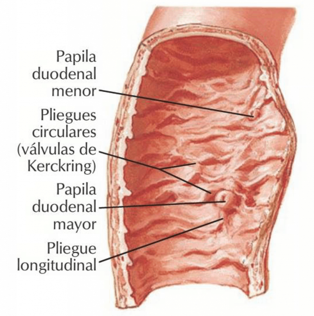
The anatomical structure of the duodenum is similar to that of the rest of the small intestine and will be studied together with that of the jejunum and ileum. However, some features of its own on its internal surface are worth noting, namely two raised formations called the duodenal papillae, where the bile duct (common bile duct) and the pancreatic ducts open, and which lie in the posteromedial wall of the 2nd, or descending, part of the duodenum.
The major duodenal papilla or caruncle lies inferiorly; it is the prominence of the hepatopancreatic ampulla (of Vater), and the main pancreatic duct (of Wirsung) reaches it together with the common bile duct.
The minor duodenal papilla or caruncle lies above the major one and the accessory pancreatic duct (of Santorini) opens into it.
Pancreas
The pancreas is a mixed gland, with an exocrine, digestive secretion (pancreatic juice) that is poured into the duodenum through the pancreatic ducts, and an endocrine secretion (glucagon and insulin) that is released into the blood and plays an essential role in carbohydrate metabolism.
This viscus is framed by the duodenum, lies behind the stomach, is attached to the prevertebral posterior wall (it is secondarily retroperitoneal) and lies in both the supramesocolic and inframesocolic regions.
External configuration
It is an elongated gland, flattened anteroposteriorly, lying more or less transversely between the duodenum on the right and the spleen on the left. It moulds itself to the lumbar spine (L1-L2) and is concave posteriorly.
It has a head, a neck, a body and a tail.
- The head of the pancreas is framed by the duodenum. It has a hook-shaped extension (the uncinate process, or hook of the pancreas), which is separated from the head of the pancreas by a notch through which the superior mesenteric vessels pass.
- The neck of the pancreas joins the head to the body. It is a somewhat narrow portion.
- The body of the pancreas extends to the left, crossing the aorta and the vertebral column.
- The tail of the pancreas is the narrower left end of the pancreas, directed towards the hilum of the spleen. It is thick and may end in a point or bluntly.
Overall, the pancreas has two surfaces: anterior and posterior; and two borders: superior and inferior.

In exceptional cases the pancreatic bud develops abnormally and may completely surround the 2nd part of the duodenum, which is called annular pancreas and may cause duodenal obstruction. On other occasions a divided pancreas, or even isolated pancreatic islets in aberrant positions, may be found.
Structurally, the gland is made up of two different tissues:
The gland of external secretion
It has pancreatic glandular acini, similar to those of the salivary glands, with excretory ducts for the pancreatic juice, which finally drain into the main and accessory pancreatic ducts, which open into the second part of the duodenum.
The gland of internal secretion
It is formed by the pancreatic islets (of Langerhans), more or less spherical in shape, lying between the acini and made up of different cell types. The cell clusters are intermingled with a vascular network and their secretory products are sent into the blood.
The pancreatic excretory ducts
There are two ducts, one main and one accessory.
The main pancreatic duct (of Wirsung)
It originates in the tail and runs along the whole length of the pancreas to the head, receiving numerous tributaries from the tail, the body and the posterior part of the head of the pancreas along its course. Near the duodenum it bends slightly until it reaches the common bile duct, and both end in the hepatopancreatic ampulla (of Vater). This opens into the duodenum at the major duodenal papilla. The hepatopancreatic ampulla is surrounded by a smooth muscle structure, the sphincter of Oddi, which controls the flow of bile and pancreatic juice into the duodenum.

The accessory pancreatic duct (of Santorini)
It drains only the anterior part of the head of the pancreas. It separates from the main pancreatic duct at the level of the head and runs transversely to the right. It ends by opening into the duodenum, 2 or 3 cm above the main pancreatic duct, at the minor duodenal papilla.
If the hepatopancreatic ampulla is blocked (by a gallstone or by cancer of the head of the pancreas), the pancreas is likely to become inflamed (pancreatitis) and bile may reflux into the pancreatic duct, which is another cause of pancreatitis.
In injuries to the pancreas caused by a sudden, forceful compression of the abdomen (as occurs in car accidents when the abdomen strikes the steering wheel), the pancreas generally ruptures as it is crushed against the vertebral column. Rupture of the pancreas tears the ducts and allows pancreatic juice to escape into the surrounding tissues, so that the pancreatic tissue and other tissues are digested, which is a serious and very painful process.

Peritoneum of the duodenopancreatic complex
The duodenopancreatic complex is covered by parietal peritoneum only on its anterior surface and is fixed to the posterior abdominal wall by a fusion fascia, Treitz's fascia (the duodenum and pancreas are secondarily retroperitoneal organs), with two exceptions, two parts of the complex that are intraperitoneal:
- The first part, which is enveloped by the peritoneum extending from the stomach. The peritoneum covering the first part of the duodenum in front and behind continues upwards with the lesser omentum and downwards with the greater omentum.
- The tail of the pancreas, which is enveloped by peritoneum, a peritoneal fold coming from the splenic hilum (pancreaticosplenic epiploon).
In addition, following the anterior surface of the duodenum and pancreas, the attachment of the transverse mesocolon can be seen crossing it obliquely, drawing a line that passes over the anterior surface of the second part of the duodenum, the head, and the lower border of the body of the pancreas. The complex is thus divided into two parts: a supramesocolic portion and an inframesocolic portion.
Relations of the duodenopancreatic complex
The duodenopancreatic complex occupies a deep position in the abdomen, inaccessible to palpation, as these are secondarily retroperitoneal organs.
Posterior relations
Through Treitz's fascia, it is related on either side to the kidneys and their pedicles and, in the centre, to the great vessels: the aorta and inferior vena cava.
Anterior relations
- Supramesocolic relations: the duodenum is related to the liver and gallbladder, and the pancreas, through the peritoneum (omental bursa), is related to the posterior surface of the stomach.
- Inframesocolic relations: they are related to the transverse mesocolon and transverse colon, the root of the mesentery, intestinal loops and the superior mesenteric vessels.