Down syndrome is a genetic disorder caused by an extra copy of chromosome 21 (a whole or partial copy, which is why the disease is known as trisomy 21).
Some of its clinical manifestations are cognitive disability, specific recognisable physical and facial features, association with organic disease (mainly cardiac, digestive and endocrine) and other abnormalities that we will study later.
The frequency of this disease is 1 in every 700 children born. It is the leading cause of severe intellectual disability in developed countries and is of great interest because genetic counselling is essential.
Remember that this lesson is part of the free courses from Paradigmia. Access the full course below 👇👇 PAEDIATRICS COURSE
Pathogenesis of Down syndrome
The mechanisms by which trisomy 21 occurs are currently unknown. Meiotic non-disjunction occurs in 96% of cases, most often in the mother (80%).
Karyotypes in Down syndrome
There are different karyotypes through which this trisomy arises. The end result is an excess of chromosome 21 (a whole duplicated chromosome or part of one).
- Free trisomy of pair 21:
- 93% of cases of Down syndrome have this karyotype.
- It is an alteration that affects all the cells of the individual’s body.
- The cause is maternal non-disjunction in 80%.

- Genetic mosaicism 21:
- 2.5% of cases of Down syndrome have this karyotype.
- This karyotypic alteration does not affect all cells.
- Clinically the syndrome is milder and less severe than a free trisomy of chromosome 21 (the previous case).


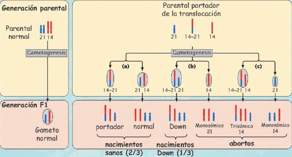
- Robertsonian translocation (qualitative):
- This karyotype is present in 4% of cases of Down syndrome.
- It is an inherited alteration and usually occurs in young mothers.
- The Robertsonian translocations in order of frequency are:
- 21-->14: the most common.
- 21-->22.
- 21-->21.
Risk factors for Down syndrome in children
- The risk increases with maternal age (a closely related factor):
- Other factors: irradiation, cytostatic drugs
- Mother’s age and frequency of Down syndrome:
- 20 years: 1/1,527
- 45 years: 1/23.

- Infertility: it is forced by hormonal treatments
Aetiology of Down syndrome
Genes related to Down syndrome (genes that are overexpressed because chromosome 21 is affected)
- Superoxide dismutase (SOD1): synthesises an enzyme that gives rise to oxygen free radicals, which increase the oxidative capacity of cells and produce premature ageing.
- ETS-2: causes leukaemias or other tumours.
- GIRK-2, KIRA: causes increased potassium efflux through cell channels and neuromuscular abnormalities responsible for hypotonia.
Remember that this lesson is part of the free courses from Paradigmia. Access the full course below 👇👇 PAEDIATRICS COURSE
Clinical manifestations of Down syndrome
Manifestations in the neonatal period
To identify it, the HALL CRITERIA must be met (especially in cases of mosaicism); otherwise the manifestations are so clear that they do not need to be met.

- Flattened facial profile.
- Excess skin on the neck.
- Upward slanting of the palpebral fissures, with the lateral canthus higher.
- Clinodactyly of the 5th finger. (abnormal deviation)
- Marked muscular hypotonia; if there is no hypotonia, doubt the diagnosis of Down syndrome. A normal newborn has a certain degree of hypertonia.
- Absent Moro reflex.
- Open mouth because of the macroglossia they usually have.
- Cheerful children, although some have autistic features; this is not the most common picture.
- Moderate intellectual disability on average.

When these signs appear we must suspect Down syndrome and perform a karyotype.
Manifestations in the older child
- Intellectual disability with hypotonia (17%): with speech difficulties and premature ageing.
- Particular phenotype: skull, face, thorax, abdomen, limbs, skeleton.
- Endocrine and metabolic abnormalities: obesity, hyperglycaemia, short stature, hypothyroidism…
- Congenital malformations: cardiac, digestive, renal, neurological… (these are the ones that lead to death).
Remember that this lesson is part of the free courses from Paradigmia. Access the full course below 👇👇 PAEDIATRICS COURSE
Morphological abnormalities caused by Down syndrome
- Growth retardation.
- Abnormalities of the face and skull:
- Brachycephaly and broad forehead.
- Flat, broad nose, sometimes even with no nasal septum.
- Small palpebral fissures, slanting outwards (almond-shaped eyes).
- Epicanthus: skin fold at the inner corner of the eye.
- Brushfield spots: small greyish spots on the iris.
- Large tongue, geographic or scrotal (as if with relief), constantly protruding because of the smallness of the mouth rather than macroglossia (sucking sign).
- Low-set, dysmorphic ears.
- Short neck with excess skin.
- Hypertelorism.
- Abnormalities of the thorax: pectus carinatum due to sternal protrusion (not common).
- Abnormalities of the abdomen:
- Abdominal hypotonia.
- Frequent umbilical hernias.
- Abnormalities of the upper limbs:
- Short, stubby fingers.
- Clinodactyly: inward curving of the 5th finger.
- Single crease across the four fingers: simian crease. It can appear in normal people.
- Abnormalities of the lower limbs:
- Increased gap between the first and second toes, which is called the sandal gap sign.
- Short, stubby toes.
- Skeletal abnormalities: they occur mainly at two levels:
- Hip: with obliquity of the acetabula (the acetabular angle is reduced).
- Atlanto-axial subluxation: can lead to myelopathy through spinal cord compression.
Congenital malformations in Down syndrome
Brain manifestations (neurological problems)
- Epilepsy in 5-10%. West syndrome.
- Vascular abnormalities, generally in the form of haemorrhage from aneurysms of the circle of Willis.
- Hydrocephalus.
- Atlanto-axial subluxation causing stenosis of the spinal canal at the high cervical level, which produces spinal cord compression and leads to myelopathy. In these children the neck must be examined carefully, and if they undergo surgery, the anaesthetist must be warned to be careful because hyperextension can cause spinal cord compression.
Manifestations of the digestive system
- Gastro-oesophageal reflux due to the frequent hypotonia of the cardia.
- Duodenal atresia.
- Aganglionic megacolon.
- Coeliac disease.
Manifestations of the digestive system
Congenital heart disease: (most common)
- Endocardial cushion defects.
- Tetralogy of Fallot. Based on ventricular septal defect, narrowing of the pulmonary artery, thickening of the muscular wall of the right ventricle (boot-shaped heart on X-ray) and overriding aorta. The most striking clinical feature of this congenital heart disease is cyanosis.

Manifestations of the respiratory system
Obstructive sleep apnoea leading to pulmonary hypertension. They are usually drowsy.
Other manifestations
- Blood: higher incidence of leukaemia (generally myeloblastic, but also lymphoblastic. Very serious).
- Endocrine: thyroid abnormalities (hypothyroidism), tendency to obesity.
- HYPOTONIA.
- Orthopaedic: hip dysplasia.
- Psychiatric: autistic features: they are not very evident; in fact, children with Down syndrome are usually very sociable and affectionate.
- Malabsorption syndrome: coeliac disease, reduced vitamin A.
- Decreased Zn, Fe and Mg and increased Ca and Cu.
- Immunological abnormalities: greater susceptibility to infections. They form abscesses more easily (they have impaired phagocytosis by macrophages).
- In women fertility is preserved, whereas in men it almost always causes infertility.
- Skin: xeroderma, alopecia areata…
- Risk of Alzheimer’s disease.
- Premature ageing: ageing in people with Down syndrome begins at around 35-45 years. By 50 years they are elderly (shortened life expectancy: 50 – 55 years).
- Eyes: keratoconus, cataracts (3%).
Remember that this lesson is part of the free courses from Paradigmia. Access the full course below 👇👇 PAEDIATRICS COURSE
Diagnosis of Down syndrome
We can distinguish the diagnosis of Down syndrome at different stages of life:
Postnatal diagnosis of Down syndrome
A suspected diagnosis is reached by physical examination, on observing the phenotype described above. It is confirmed by karyotype and banding techniques.
Prenatal diagnosis of Down syndrome in the 2nd trimester of pregnancy
The diagnosis is made using non-invasive tests on maternal blood:
- Triple test: reliability 70%. ↓α-fetoprotein, ↑β-gonadotropin, ↓unconjugated oestradiol.
- Quadruple test: reliability 80%. α-fetoprotein, β-gonadotropin, unconjugated oestradiol, inhibin protein.

- Ultrasound: reliability 95%. The nuchal fold and absence of the nasal bone are identified.

To confirm the diagnosis, invasive tests must always be done:
- Amniocentesis, chorionic villus sampling: reliability 100%.
- Karyotype: from the samples obtained in the previous tests. If the karyotype is normal, we do FISH. Reliability 100%.
There may be mosaicisms that are not so apparent and are therefore not detected.
Clinical prognosis of Down syndrome
Reduced life expectancy (they live up to 50-55 years) due to:
- Heart disease.
- Digestive abnormalities (oesophageal atresia associated with fistulas, duodenal atresia, aganglionic megacolon).
- Haematological problems: acute lymphoblastic leukaemia (97% of cancers in children with Down syndrome).
- Alzheimer’s disease.
- Pulmonary hypertension due to sleep apnoea.
- Infections.
- Seizures.
Survival of patients with Down syndrome
- 75% of fetuses suffer intrauterine death.
- Of those born: 15% die before the age of 1 year, 35% die before the age of 50 years, 50% survive beyond 50 years.
- In 1960, survival was only 12 months; in 2005 it was 50-55 years.
Remember that this lesson is part of the free courses from Paradigmia. Access the full course below 👇👇 PAEDIATRICS COURSE
Prevention of Down syndrome
- Genetic counselling: taking into account maternal age and the karyotype of the parents if there is a family history, and above all if they are under 35 years.
- Prenatal diagnosis.
In the case of the karyotype of young parents:
- If the child with Down syndrome has a translocation and the parents have a normal karyotype, the incidence of another child with Down syndrome is normal.
- If the father has a 21-->14 translocation and the mother a normal karyotype, the incidence does not increase.
- If the father has a normal karyotype and the mother a 21-->14 translocation, the incidence increases.
- If the father has a 21-->22 translocation and the mother a normal karyotype, the incidence changes little.
- If the father has a normal karyotype and the mother a 21-->22 translocation, the probability will be 33%, that is, high.
- If the father has a 21-->21 translocation and the mother a normal karyotype, the probability is 100%.
- If the father has a normal karyotype and the mother a 21-->21 translocation, the incidence will also be 100%.
Recommended reading:
- Síndrome de down (information for parents)
- Síndrome de down (information for children)
- The neonate or newborn
- Síndrome de Down: MedlinePlus en español