In this lesson of the MUSCULOSKELETAL SYSTEM COURSE we will look at the characteristics of synovial joints and the types that exist.
As we saw in the joints module, there are several types of joints. One of these types is synovial joints.
Synovial joints are connections between skeletal components in which the elements involved are separated by a narrow joint cavity.
As well as having this joint cavity, synovial joints have several characteristic features.
First, they have a layer of cartilage (usually hyaline cartilage). This layer covers the articular surfaces of the skeletal elements so that the bony surfaces do not come into direct contact with each other.
As a result, when these joints are seen on plain radiographs, a wide gap appears to separate the adjacent bones, because the cartilage covering the articular surfaces is more transparent to X-rays than bone. Let us see this in the following image:

Remember that this lesson is part of the free courses from Paradigmia. Access the full course below 👇👇 MUSCULOSKELETAL SYSTEM COURSE
Synovial joints are all the joints of the limbs except the tibiofibular joint.
Elements of synovial joints

1. Cavity
It is closed and filled with synovial fluid.
2.Articular surfaces
These are the part of the bony end that comes into contact with the other bone.
They are lined with articular cartilage, which is hyaline cartilage in most cases, except in the temporomandibular and sternoclavicular joints, where it is fibrocartilage.
3. Fibrous capsule and ligaments
This is what closes the synovial joint, and it is reinforced by ligaments and by the tendons of the muscles surrounding the joint. It encloses the bony ends that come into contact. It is the main means of union of the joint, preventing the ends from separating. The ligaments that reinforce the capsule can be of different types:
- Intrinsic ligaments: those found within the thickness of the capsule itself. E.g. the hip joint.
- Extrinsic extra-articular ligaments: ligaments independent of the capsule, lying outside it and outside the joint.
- Extrinsic intra-articular extrasynovial ligaments: these are like the previous ones, but in this case they lie inside the joint without being bathed in synovial fluid. Like the cruciate ligaments of the knee.
4. Synovial membrane
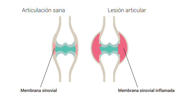
This secretes synovial fluid, produced by synoviocytes (cells), and lines the fibrous capsule on the inside and the part of the bone that is not covered by articular cartilage. It is the nutritive source of the articular cartilage and is highly vascularised. The functions of synovial fluid are nutrition, lubrication and removal of debris.

Arthritis is an acute process of inflammation of the synovial membrane. Furthermore, because the membrane is vascularised, if a vessel ruptures and blood is released into the joint, what is known as haemarthrosis (accumulation of blood in the joint) occurs.
Synovial bursae or serous bursae
Synovial or serous bursae are cavities that surround a space containing synovial fluid, which may or may not be in contact with the surface of the joint.
They can be glove-like evaginations of the synovial membrane (communicating with the joint) or they can lie outside the joint cavity like a cushion. Inflammation of the serous bursae is called bursitis. The function of these bursae is to reduce the friction, rubbing or wear of the tendons, facilitating the gliding of the muscles. E.g. the femur.

All synovial joints are diarthroses and all diarthroses are synovial joints, but when we talk about diarthroses we are referring to their function and when we talk about synovial joints we are referring to their structure.
Remember that this lesson is part of the free courses from Paradigmia. Access the full course below 👇👇 MUSCULOSKELETAL SYSTEM COURSE