In this lesson of the MUSCULOSKELETAL SYSTEM COURSE we will look at the characteristics of solid joints.
Remember that this lesson is part of the free courses from Paradigmia. Access the full course below 👇👇 MUSCULOSKELETAL SYSTEM COURSE
As we saw in the joints module, there are several types of joints. One of these types is solid joints, which are divided into:
- Fibrous joints
- Cartilaginous joints
- And finally, bony joints
We can see this better in the following image:

1. Fibrous joints
In fibrous joints the bones are united by fibrous tissue. Fibrous joints include sutures, gomphoses and syndesmoses.
1.1 Suture
This subtype of fibrous joint is found only at the junctions between the bones of the cranial vault.
We can distinguish between:
- Plane sutures: unions between smooth surfaces. Example: the nasal bones

- Serrate (dentate) sutures: where the edges interdigitate. Example: the sagittal suture.

- Bevelled suture: where the bones overlap. Example: between the temporal and parietal bones.

- Schindylesis: E.g. the inferior crest of the sphenoid and the vomer: the crest of the body of the sphenoid fits into the groove of the vomer.
All sutures are functionally immobile: synarthroses. (No movement occurs between the articulated elements)
Finally, let us look at this summary image from VisibleBody
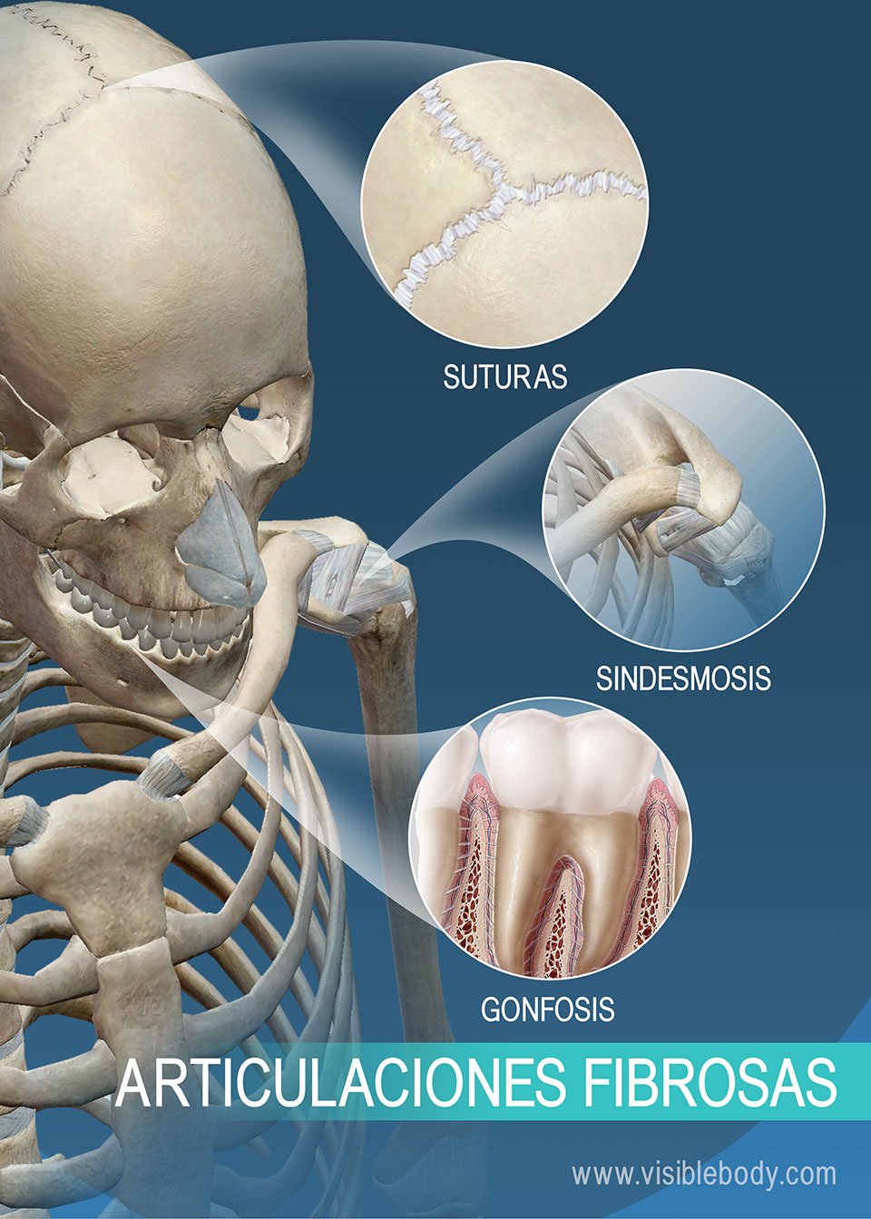
1.2 Gomphosis
They are found only between the teeth and the alveolus. In these joints the periodontal ligament joins the tooth roots to the alveoli. Functionally they are synarthroses. (No movement occurs between the articulated elements)

1.3 Syndesmosis
In these there is a wide area of fibrous tissue in the form of a membrane or interosseous ligament. Example: the distal tibiofibular joint. All the joints of the limbs have synovial cavities; the only one that is not synovial is the tibiofibular joint.
Functionally they are amphiarthroses. (Movement does occur between the articulated elements, over a small range)

2-Synovial joints
Synovial joints are those in which the bones are united by cartilage. They include synchondroses and symphyses:
2.1. Synchondrosis
They are formed by hyaline cartilage.
E.g. the following are synchondroses: the growth cartilage of bones (metaphysis); in children, the hip bone (ilium, ischium and pubis) is joined by triradiate cartilage; the body of the sternum, which is formed by bones joined by growth cartilage that gradually closes; and the sphenobasilar joint.
They are transient joints and synarthroses.
2.2 Symphysis
They occur between two bones joined by fibrocartilage.
Examples: the pubic symphysis, the joints between vertebral bodies, the mental symphysis (in children).
Between them there is relative movement: amphiarthrosis.

2.3 Bony joints
These joints consist of bones joining bones.
Example: the sacrum is formed by five bones (5 sacral vertebrae) that are separate.
They are synarthroses.
Remember that this lesson is part of the free courses from Paradigmia. Access the full course below 👇👇 MUSCULOSKELETAL SYSTEM COURSE
Some links of interest to complement your study: