The classical nomenclature used to be the Basel Nomina Anatomica (1895); before that, each country used its own nomenclature.
Today, however, the international Paris nomenclature (1895) is used:
- Latin, adapted to the vernacular language.
- A single name that is short, simple and descriptive.
- Structures that are topographically related have similar names.
- Eponyms (proper names) are not used.
Anatomical position
Standing upright, looking straight ahead, with the arms at either side of the trunk. The palms of the hands face forwards, with the forearm supinated. (axes missing)
Planes:
- Transverse or horizontal.
- Median sagittal or anteroposterior (if it is not in the midline it is called parasagittal)
- Coronal or frontal
Sections:
- Transverse or horizontal.
- Median sagittal or anteroposterior.
- Coronal or frontal.

Terms of location or position:
- Proximal: closer to the root of the limb.
- Distal: further from the root of the limb.
- Ulnar or medial
- Radial, lateral or external.
- Plantar: the sole of the foot
- Dorsal: the dorsum of the foot.
- Palmar: the palm of the hand.
- Dorsal: the dorsum of the hand.
- Homolateral or ipsilateral: on the same side.
- Heterolateral or contralateral: on the opposite side.

Terms of movement:
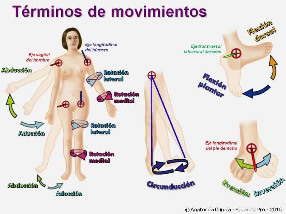
There are three degrees of freedom of movement; all other movements are combinations of these three:
1. Flexion and extension
This movement takes place around a transverse axis and occurs in a sagittal plane. Flexion is generally regarded as bringing the anterior surfaces closer together, and extension as bringing the posterior surfaces closer together or moving the anterior ones apart.
One of the exceptions to this general rule is the knee, where flexion brings the posterior surfaces closer together. In the foot, dorsiflexion brings the anterior surfaces closer together (when walking), whereas plantar flexion brings the posterior surfaces closer together (standing on tiptoe). Lateral flexion can also occur in the trunk.
2. Moving apart and bringing together (abduction and adduction)
This occurs around a sagittal axis and in a coronal plane. In the hand, the anatomical position must be taken into account for these movements, since there is an ulnar or medial movement (moving apart) and a lateral or radial one (bringing together).
3. Rotation
This occurs around a vertical axis and in a transverse plane. Rotation of the trunk is described from right to left, whereas in the limbs it is described from medial (internal) to lateral (external).
The hands are in lateral rotation in supination and in medial rotation in pronation. The feet can be inverted, with the sole turned inwards, or everted, with the sole turned outwards.
4. Circumduction
All three movements at once.
Huesos de la rodilla derecha
Ver en el atlas de cuerpo completo ↗
Activa el modelo para explorar sus estructuras.
Modelo anatómico real de referencia. Selecciona una estructura para destacarla o aislarla.
Objetivo: Identificar el fémur, la tibia, la fíbula y la rótula derechas, y reconocer su relación espacial en la región de la rodilla.
Descripción y licencia
Vista tridimensional de los huesos de la rodilla derecha. El extremo distal del fémur se sitúa sobre la tibia; la fíbula queda en el lado lateral y la rótula está por delante de la articulación. El modelo muestra únicamente huesos y no incluye cartílago, meniscos ni ligamentos.
BodyParts3D, © The Database Center for Life Science · CC BY 4.0
Modelo educativo de referencia. No es una herramienta de diagnóstico. Se muestran las etiquetas del catálogo curricular; la terminología generada automáticamente no se publica desde este visor.