You can study all these muscles in greater depth in the Paradigmia® muscle tables
The tendons that run from the forearm to the fingers glide, at the wrist and in the fingers, through fibrous canals surrounded by synovial sheaths.
1. Anterior region of the wrist: flexor retinaculum
The superficial fascia of the forearm thickens at the wrist and becomes a thick ligament at the level of the carpal bones; this ligament is called the flexor retinaculum or transverse carpal ligament.

This ligament is attached at four bony points: the pisiform, the hook of the hamate bone, the tubercle of the scaphoid and the tubercle of the trapezium. In this way the flexor retinaculum converts the groove of the carpal bones into a canal called the carpal tunnel (carpal canal) through which the flexor tendons run to the hand, except that of pronator teres, which does not reach it, and palmaris longus, which passes over it.
From the deep surface of the transverse carpal ligament a fibrous septum arises that divides it into two canals, a lateral one for flexor carpi radialis and a medial one for the tendons of flexor digitorum superficialis, flexor digitorum profundus and flexor pollicis longus, enclosed in their synovial sheaths, together with the median nerve.
In addition, on its superficial surface the flexor retinaculum receives a fibrous expansion from the tendon of flexor carpi ulnaris, which delimits a tunnel, Guyon’s canal (the ulnar canal), through which the ulnar nerve and vessels and the tendon of flexor carpi ulnaris pass.
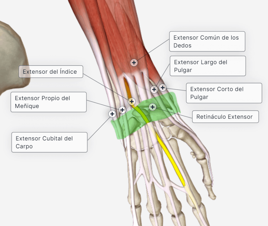
2. Posterior region of the wrist: extensor retinaculum
It is also a continuation of the superficial fascia of the forearm, which thickens and forms the extensor retinaculum or dorsal carpal ligament that attaches laterally to the triquetrum and pisiform and to the distal end of the radius; numerous septa arise from its deep surface and attach to the margins of the grooves on the posterior surface of the ulna and radius, thus delimiting a series of osteofibrous canals or tunnels through which the tendons run.
There are a total of six separate tunnels, which, from the radial to the ulnar border, correspond to the following tendons, enclosed in their synovial sheaths:
- Abductor pollicis longus and extensor pollicis brevis
- Extensor carpi radialis longus and brevis
- Extensor pollicis longus
- Extensor digitorum and extensor indicis
- Extensor digiti minimi
- Extensor carpi ulnaris
3. Synovial sheaths of the flexor tendons
To facilitate the gliding of the flexor and extensor tendons of the fingers through the osteofibrous tunnels. There are dorsal and palmar synovial sheaths.
Ganglion: a benign cystic tumour with a more or less fluid content, found mainly on tendons or aponeuroses of the wrist or on the dorsum of the foot, and formed by a thin fibrous capsule with mucoid contents.

4. Movements of the carpus
The carpus can perform flexion–extension and radial and ulnar deviation.