You can study all these muscles in greater depth in the Paradigmia® muscle tables
Scapular muscles
The scapular muscles are a group of muscles that attach to the scapula and to the proximal end of the humerus and move the glenohumeral joint (they do not move the scapula). The glenohumeral joint performs flexion–extension, adduction and abduction, and lateral and medial rotation.
The glenohumeral joint is very mobile but very unstable, so these muscles act like ligaments that hold the joint together. The scapular muscles are: subscapularis, supraspinatus, infraspinatus, teres minor, teres major and deltoid.
1. Lateral rotators
They insert on the greater tubercle of the humerus and pull it laterally, producing lateral rotation. They are infraspinatus, teres minor and the posterior part of the deltoid.
1.1 Infraspinatus
It arises together with teres minor in the infraspinous fossa; its fibres run horizontally laterally, pass behind the glenohumeral joint and insert on the greater tubercle.
- Action: When it contracts it produces adduction and lateral rotation of the arm.

1.2 Teres minor
It lies inferior to infraspinatus and, like it, arises in the infraspinous fossa, passes behind the glenohumeral joint and inserts on the greater tubercle of the humerus.
- Action: when it contracts it produces adduction and lateral rotation of the arm.
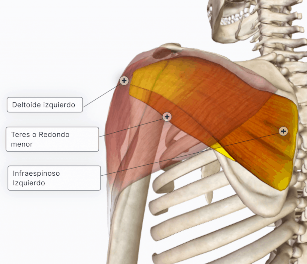
1.3 Deltoid (posterior part)

2. Medial rotators
They insert on the lesser tubercle and pull it forwards and medially. There are 4 main muscles: 2 of them belong to the scapular (periscapular) muscles: teres major and subscapularis; the other 2 are larger and lie further away: latissimus dorsi and pectoralis major.
In addition, coracobrachialis and the anterior part of the deltoid also produce this movement.
2.1 Subscapularis
It arises in the subscapular fossa of the scapula, passes in front of the glenohumeral joint and inserts by a tendon on the lesser tubercle of the humerus.
- Action: when it contracts it produces adduction and medial rotation of the arm.
2.2 Teres major
It arises from the lateral (axillary) border of the scapula, below teres minor, near the inferior angle; its fibres run upwards and laterally, passing below the joint, to insert by a tendon on the crest of the lesser tubercle of the humerus, below subscapularis.
- Action: adduction and medial rotation.
2.3 Latissimus dorsi
2.4 Pectoralis major

2.5 Coracobrachialis
It lies in the superomedial part of the arm, covered by the short head of biceps brachii. It arises from the coracoid process and runs downwards and slightly laterally, passing behind pectoralis major, to insert on the medial surface of the shaft of the humerus, at its midpoint.
- Action: when it contracts it produces flexion, medial rotation and adduction of the arm, and flexion of the elbow.

2.6 Deltoid (anterior part)

3. Abductors
3.1 Deltoid
A thick muscle shaped like a hollow half-cone, with its base above and its apex below. It lies over the lateral part of the shoulder, giving it its rounded contour. When the nerve that supplies it (the “deltoid nerve”) is injured, the muscle atrophies and the rounded contour is lost (“epaulette” shoulder).
The deltoid has 3 parts:
- Anterior or clavicular: arises from the lateral half of the clavicle.
- Middle or acromial: arises from the acromion.
- Posterior or spinal: arises from the spine of the scapula, which lies posteriorly.
These parts converge and end by inserting on the humerus at the deltoid tuberosity (deltoid V). Lateral surface of the shaft of the humerus
- Action: as a whole, the deltoid is an abductor of the arm. But if it contracts in isolation, the anterior part can produce medial rotation and flexion (like pectoralis major) and the posterior part can produce lateral rotation and extension (like latissimus dorsi). Depending on the position of the humerus, the axis of abduction lies within the deltoid, and if abduction starts from a small degree of abduction, the axis already lies below it. Depending on the position of the humerus, one part or another will be more powerful.
3.2 Supraspinatus
It arises in the supraspinous fossa, passes above the shoulder joint, below the acromion, and inserts on the greater tubercle.
- Action: when it contracts it produces abduction; it is the initiator (starter) of abduction.
If this muscle is injured, these people cannot initiate abduction of the arm but can continue it. The deltoid and supraspinatus act in a chain: supraspinatus starts the movement and the deltoid, which is more powerful, completes it.
Clinical note
The painful arc (impingement syndrome): the supraspinatus tendon is compressed because of a degenerative process, which can lead to a rotator cuff tear and even to glenohumeral osteoarthritis.
4. Rotator cuff
A group of 4 muscles that surround the shoulder joint like a cuff and are rotators: supraspinatus, infraspinatus, teres minor and subscapularis.
Their function is to stabilise the joint, and they rotate it both medially and laterally.
They are as follows:
- Supraspinatus: runs from the medial border of the scapula to the greater tubercle. → Initiates abduction
- Infraspinatus: runs from the medial border of the scapula to the greater tubercle. → Adductor and medial rotator
- Teres minor: runs from the infraspinous fossa to the greater tubercle → Adductor and lateral rotator
- Subscapularis: runs from the subscapular fossa to the lesser tubercle → Adductor and lateral rotator
5. Adductors
The main ones are latissimus dorsi and pectoralis major. The force of gravity, teres major, coracobrachialis and the long head of triceps also contribute.
- Latissimus dorsi
- Pectoralis major
- Teres major
- Coracobrachialis
- Triceps (long head)
6. Flexors (anteversion)
These muscles pass in front of the joint. The main ones are pectoralis major and the deltoid. Coracobrachialis and biceps brachii are also flexors, but less important.
- Pectoralis minor (clavicular and sternoclavicular parts)
- Deltoid (anterior or clavicular part)
- Coracobrachialis
- Biceps brachii
7. Extensors (retroversion)
They pass behind the joint. The most important are teres major, latissimus dorsi and the posterior part of the deltoid. Triceps also contributes.
- Teres major
- Latissimus dorsi
- Deltoid (posterior or spinal part)
- Triceps
