1. Muscles of the cervical region
In the neck there are dorsal, lateral and prevertebral muscles. The nuchal septum divides the musculature of the nape into a right half and a left half.
1.1 Dorsal muscles
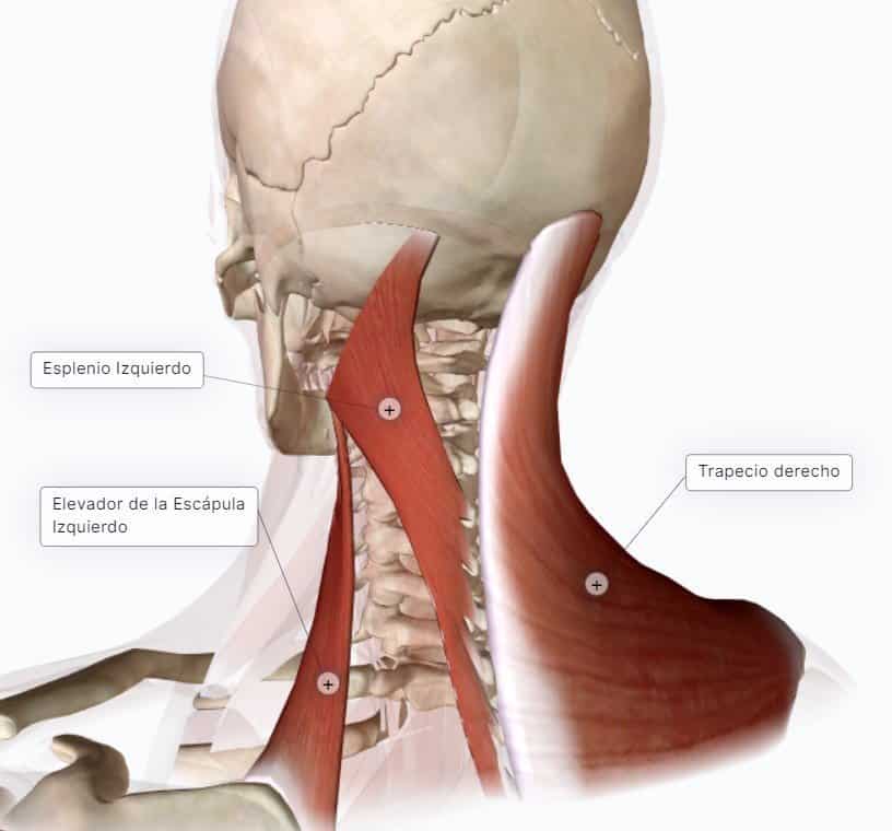
Beneath the subcutaneous cellular tissue lies trapezius (descending fibres), which produces extension, lateral flexion and contralateral rotation of the head. Next, beneath trapezius, lie splenius and, laterally, levator scapulae.
1.1.1 Splenius
It lies deep to trapezius and medial to levator scapulae. It is enclosed by an expansion of the fascia. It has two parts, splenius capitis and splenius cervicis.
- Splenius capitis originates from the spinous processes of the last cervical and first thoracic vertebrae, and its fibres ascend to insert on the mastoid process and below the superior nuchal line.
- Splenius cervicis originates from the spinous processes of the thoracic vertebrae and ends on the transverse processes of the cervical region (without reaching the head).
Action:
- Extension of the head (it draws it backwards).
- Lateral flexion of the head
- Ipsilateral rotation of the head: so that if the left splenius contracts, the head turns to the left and vice versa (fibres running upwards and medially produce contralateral rotation, like trapezius; and those running upwards and laterally, like splenius, produce ipsilateral contractions).
1.1.2 Semispinalis capitis, or complexus major
It lies beneath splenius. It is the most cranial part of the transversospinal system and runs from the transverse processes of the upper thoracic and cervical vertebrae to insert on the occipital bone, between the superior and inferior nuchal lines. Its most important action is extension of the head (antigravity). It can carry out a small rotation of the head, but this is of little importance.
Beneath splenius lie, from medial to lateral: semispinalis capitis, longissimus capitis or complexus minor (the most cranial part of longissimus), longissimus cervicis and, most laterally, iliocostalis cervicis.
1.1.3 Short muscles of the head, or suboccipital muscles
They lie beneath the anterior muscles, in the deepest plane of the dorsal surface. There are four muscles, lying against the craniovertebral complex. These muscles move the head. They lie in a limited space, bounded superiorly by the occipital bone (inferior nuchal line), the posterior arch of the atlas and the arch of the axis with its spinous process.

From medial to lateral they are:
- Rectus capitis posterior minor: the most medial. It runs from the posterior tubercle of the atlas to the most medial part of the inferior nuchal line. This muscle produces extension of the head.
- Rectus capitis posterior major: runs from the spinous process of the axis to the most lateral part of the inferior nuchal line; it can produce lateral flexion and extension, and, unilaterally, ipsilateral rotation.
- Obliquus capitis superior (minor): runs from the transverse process of the atlas and inserts between the superior and inferior nuchal lines, above rectus capitis posterior major. It produces extension of the head and, if it contracts unilaterally, lateral flexion.
- Obliquus capitis inferior (major): from the spinous process of the axis to the transverse process of the atlas. It produces ipsilateral rotation and lateral flexion.
Beneath the occipital bone, and related to the suboccipital muscles, lies the suboccipital triangle. Its boundaries are obliquus capitis inferior, obliquus capitis superior and rectus capitis posterior major. At the floor of the triangle lie the posterior arch of the atlas, the posterior atlanto-occipital membrane, the vertebral artery and the dorsal ramus of the 1st cervical spinal nerve.
This ramus has its own name, suboccipital nerve (dorsal ramus of the 1st cervical spinal nerve), which provides the motor innervation of the suboccipital muscles. It has no sensory distribution; the sensory innervation of this area is provided by the greater occipital nerve, the dorsal ramus of the 2nd cervical spinal nerve.
The muscles of the back are innervated by the dorsal rami of the spinal nerves, depending on the level at which they lie (cervical, thoracic, lumbar, sacral).
