The innervation of the lower limb comes from two nerve structures known as the lumbar plexus and the sacral plexus. We will study their anatomy, the possible pathologies of the sacral plexus, its course and the nerves that make it up. We will also look at its most important branches, such as the sciatic nerve, the pudendal nerve and the posterior femoral cutaneous nerve.
Remember that this lesson is part of the free courses from Paradigmia. Access the full course here 👉👉 MUSCULOSKELETAL SYSTEM COURSE
- What is the sacral plexus?
- Collateral branches of the sacral plexus
- Terminal branches
- 1- Superior gluteal nerve (L4-S1)
- 2- Inferior gluteal nerve (L5-S2)
- 3- Posterior femoral cutaneous nerve (S1-S3)
- 4- Pudendal nerve (S2-S4)
- 5- Sciatic (ischiadic) nerve (L4-S3)
- 6- Tibial nerve (L4-S3)
- 7- Common fibular nerve (L4-S2)
- Recommended reading on the lumbar plexus
What is the sacral plexus?
The sacral plexus is formed by the lumbosacral trunk (made up of the anterior ramus of L5 plus an anastomotic branch from L4) and by the anterior sacral nerves from S1 to S4.
On its course it leaves the pelvis resting on the piriformis muscle. The sacral plexus gives a series of branches to the lower limb.

Collateral branches of the sacral plexus
These are short branches that stay in the vicinity of the plexus and supply the pertrochanteric muscles, so they are motor branches. The pertrochanteric muscles are lateral rotators of the hip joint and are: the piriformis, the superior and inferior gemelli (spinal and tuberal), the obturator internus and externus, the quadratus femoris, etc.

Terminal branches
These are longer branches that leave through the greater sciatic foramen above or below the piriformis muscle. They are:
- Superior gluteal n.
- Inferior gluteal n.
- Posterior femoral cutaneous n.
- Pudendal n.
- Sciatic n.
1- Superior gluteal nerve (L4-S1)
The superior gluteal nerve leaves the pelvis through the suprapiriform foramen (through the greater sciatic foramen above the piriformis muscle), together with the superior gluteal artery. It gives motor branches to the gluteus medius, gluteus minimus and tensor fasciae latae.

2- Inferior gluteal nerve (L5-S2)
The inferior gluteal nerve leaves the pelvis through the infrapiriform foramen (the greater sciatic foramen below the piriformis muscle), together with the inferior gluteal artery, and is distributed to the gluteus maximus, providing its motor supply.
Both the superior and inferior gluteal nerves, like the collateral branches, have only a motor distribution.

Remember that this lesson is part of the free courses from Paradigmia. Access the full course here 👉👉 MUSCULOSKELETAL SYSTEM COURSE
3- Posterior femoral cutaneous nerve (S1-S3)
The posterior femoral cutaneous nerve leaves through the greater sciatic foramen, via the infrapiriform foramen, medial to the sciatic nerve, resting on the pertrochanteric muscles. It is distributed over the whole posterior aspect of the thigh and the popliteal fossa, supplying these areas sensorily. It has no motor territory (it is a cutaneous nerve).
The posterior femoral cutaneous nerve gives a series of branches:
- Perineal branches: go to the posterior part of the perineum, supplying it sensorily.
- Inferior cluneal nerves: supply the lower part of the buttock sensorily.
The sensory supply of the buttock is as follows:
- The upper part is supplied sensorily by the superior cluneal nerves that come from the dorsal rami of L1 to L3.
- The medial part is supplied sensorily by the medial cluneal nerves that come from the dorsal rami of S1 to S3.
- The lateral part is supplied by the lateral cutaneous branch of the iliohypogastric nerve (T12-L1) (lumbar plexus).
- The lower part is supplied sensorily by the inferior cluneal nerves, branches of the femoral cutaneous nerve (S1-S3) (sacral plexus).
The superior and medial cluneal nerves come from neither the sacral plexus nor the lumbar plexus but are the dorsal rami of the spinal nerves.

4- Pudendal nerve (S2-S4)
It leaves the pelvis through the greater sciatic foramen, via the infrapiriform foramen, and re-enters the pelvis through the lesser sciatic foramen into the ischioanal (ischiorectal) fossa.
The pudendal nerve forms a plexus that provides the motor supply to the pelvic floor and the urogenital diaphragm. It also has a sensory territory, the perineum.
The anterior part of the perineum is supplied by the ilioinguinal nerve, and the pudendal nerve collects the sensation of the posterior part of the perineum.

5- Sciatic (ischiadic) nerve (L4-S3)
The anterior rami of L4 to S3 converge in the sciatic nerve, which leaves through the infrapiriform foramen, covered by the gluteus maximus and resting on the pertrochanteric muscles.
The sciatic nerve descends through the posterior compartment of the thigh, covered by the long head of the biceps femoris and resting on the adductor magnus, which it supplies motorically (the adductor magnus is also supplied motorically by the obturator nerve from the lumbar plexus).
In this area it also gives motor branches to the three hamstrings (biceps femoris, semitendinosus and semimembranosus).
As it approaches the popliteal region it gives its two terminal branches:
- Tibial n. (classically the internal popliteal sciatic nerve → L4-S3)
- Common fibular n. (classically the external popliteal sciatic nerve → L4-S2)
The sciatic nerve also provides the motor supply to the hamstring muscles: the short head of the biceps femoris is innervated by the common fibular nerve, whereas the long head, the semitendinosus and the semimembranosus are innervated by the tibial nerve.
As the sciatic nerve, it gives no sensory branch, but its branches do have a sensory distribution.

Remember that this lesson is part of the free courses from Paradigmia. Access the full course here 👉👉 MUSCULOSKELETAL SYSTEM COURSE
6- Tibial nerve (L4-S3)
It is the more medial one. It descends through the popliteal fossa accompanying the popliteal vessels. It is the most superficial component of this neurovascular bundle (from superficial to deep: the nerve, the vein and the posterior tibial artery).
It descends through the popliteal fossa and rests on the popliteus muscle, then passes through the tendinous arch of the soleus, together with the posterior tibial artery. It descends through the whole posterior compartment of the leg between the soleus and the deep muscles of the leg, then passes behind the medial malleolus together with the posterior tibial artery, reaching the medial border of the foot, where it divides into its two terminal branches.
The tibial nerve gives:
- Motor branches: to the superficial compartment of the leg: triceps surae, popliteus and plantaris, as well as the whole deep compartment of the leg.
- Sensory branches: the anterior tibial nerve gives two sensory nerves:
- Medial sural cutaneous nerve: in the popliteal fossa. It descends superficially between the heads of the gastrocnemius, accompanying the small saphenous vein. This nerve receives a branch from the common fibular nerve, called the fibular communicating branch, and from that point the medial sural cutaneous nerve changes its name and is called the sural nerve. This continues to descend superficially to the dorsum of the foot, where it gives a series of sensory branches: the lateral dorsal cutaneous nerves that collect sensation from the most lateral part of the dorsum of the foot.
- Calcaneal nerves: at the level of the calcaneal tuberosity, supplying that area sensorily.
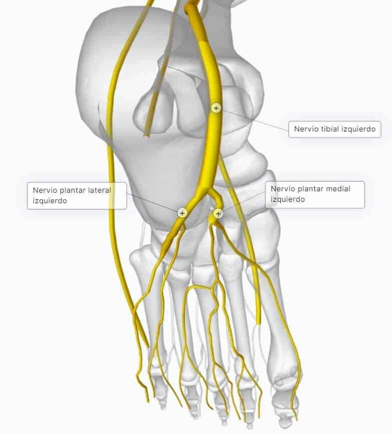
On the sole of the foot, at the level of the medial border of the foot, the tibial nerve divides into its two terminal branches, which lie deep to the flexor digitorum longus muscle:
- Medial (internal) plantar nerve: has a motor distribution, giving branches to the abductor hallucis, the medial fascicle of the flexor hallucis brevis and the first two lumbricals. It supplies the medial part of the sole of the foot sensorily.
- Lateral (external) plantar nerve: provides the motor supply to all the other muscles. It supplies the lateral part of the foot sensorily. (Sensation from the lateral border of the sole of the foot is collected by the sural nerve and that from the medial border by the saphenous nerve.)
7- Common fibular nerve (L4-S2)
The common fibular nerve runs along the most lateral and superior part of the popliteal fossa, partly covered by the biceps tendon, and then enters the fibular compartment to give its terminal branches. In a fracture of the fibula, the common fibular nerve can easily be injured.
Before this point it gives no motor branches, only two sensory branches:
- Lateral sural cutaneous nerve: a very important sensory branch that runs along the gastrocnemius and gives a series of branches that collect sensation from the posterolateral aspect of the proximal half of the leg.
- Fibular communicating branch: communicates with the medial sural cutaneous nerve to form the sural nerve.
At the level of the neck of the fibula, in the fibular compartment, it gives its two terminal branches:
- Superficial fibular nerve (musculocutaneous): descends through the fibular compartment, providing the motor supply to these muscles. It also gives cutaneous branches: to do so, at the level of the distal third of the leg it becomes superficial and heads towards the dorsum of the foot, where it gives the intermediate and lateral dorsal cutaneous nerves that are distributed to the skin of the dorsum of the foot. These branches supply the lowest and most anterior part of the leg and the dorsum of the foot, except the first interosseous space, sensorily.
- Deep fibular nerve (anterior tibial): pierces the interosseous membrane together with the anterior tibial artery, both being deep structures. It then passes beneath the extensor retinaculum and ends on the dorsum of the foot. It provides the motor supply to the whole anterolateral compartment of the leg: tibialis anterior, extensor hallucis longus, extensor digitorum longus and fibularis tertius (anterior fibular); as well as extensor digitorum brevis and extensor hallucis brevis. It also supplies the skin covering the dorsum of the first interosseous space sensorily.

Remember that this lesson is part of the free courses from Paradigmia. Access the full course here 👉👉 MUSCULOSKELETAL SYSTEM COURSE