The bones of the forearm are joined at their proximal and distal ends by two synovial joints, which act together, allowing movements of the radius on the ulna about a vertical axis (pronation-supination). They are joints of the trochoid type.
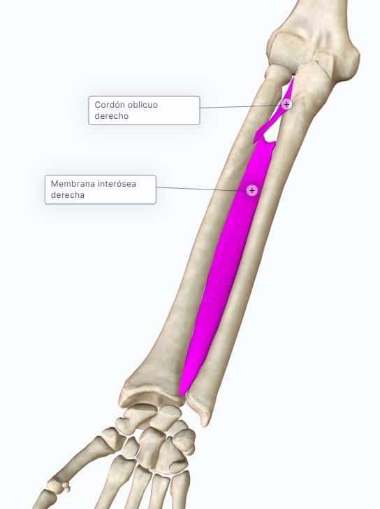
In addition, the diaphyses of both forearm bones are joined to each other by an interosseous membrane and the oblique cord.
At the distal radio-ulnar joint the distal ends of the ulna and the radius come into contact (together with an articular disc: the triangular ligament, which separates the radio-ulnar joint from the wrist joint). Functionally, this joint does not belong to the wrist.
Elements of the synovial joint
The means of union are the joint capsule and the reinforcing ligaments:
Ligaments
- Palmar or anterior radio-ulnar ligament: on the anterior part of the joint.
- Dorsal or posterior radio-ulnar ligament: behind.
- Triangular ligament: it is a sheet of fibrocartilage arranged horizontally, on the lower surface of the joint, covering the lower end of the ulna. Its apex attaches to the lateral surface of the root of the styloid process of the ulna, and its base is fixed to the lower border of the ulnar notch of the radius. Its upper surface comes into contact with the joint capsule and with the ulna, and its lower surface rests on the bones of the 1st row of the carpus. It is lined with articular cartilage, forming an articular surface for the head of the ulna (and another, inferior, for the carpal bones).
This last ligament is thinned in its central part and sometimes perforated, connecting the distal radio-ulnar joint with the radiocarpal joint.

Synovial membrane
Within the joint capsule there is a synovial membrane that is common to the double joint: that of the radius with the ulna and that of the head of the ulna with the triangular ligament. It lines the capsule internally, and in the middle and upper part it rises, forming a blind sac between the two forearm bones called the saccular recess or superior blind sac.
Interosseous membrane
The interosseous membrane is a strong fibrous sheet between the interosseous borders of the ulna and the radius. Its fibres are oblique, descending from the radius to the ulna, but in the upper part it has a free border (2-3 cm from the bicipital tuberosity of the radius). The posterior interosseous vessels pass through this gap. Distally it is continuous with the fascia of the pronator quadratus (the deepest), and below it is perforated by the anterior interosseous vessels. It not only joins but also divides the forearm into an anterior and a posterior region. It serves to increase the area of attachment of the deep muscles of the forearm.
Oblique cord or Weitbrecht's ligament
It is a more or less distinct fibrous band, sometimes double, above the interosseous membrane; it is oblique downwards and outwards. It runs from the base of the coronoid process towards the radius, below the bicipital tuberosity of the radius. It runs in a direction opposite to most of the fibres of the interosseous membrane.
Both structures (interosseous membrane and oblique cord) form the radio-ulnar syndesmosis.
Mobility
Both radio-ulnar joints are trochoid joints and act together, performing movements of pronation-supination. Pronation is limited by the quadrate ligament and the anterior radio-ulnar ligament. Supination is limited by the quadrate ligament and the posterior radio-ulnar ligament.