Here is the syllabus covered in the previous video. If you have any questions, post them in the comments section at the bottom of the page.
Protein structure of the TCR
The T-cell receptor (TCR) consists of a collection of membrane proteins, some involved in antigen recognition (variable proteins) and others with a structural function or a role in transmitting the signal into the cell (constant proteins).
Variable proteins
As already described, there are two possible T-cell receptors in terms of the composition of the variable chains: α-β (T2 cells) and γ-δ (T1 cells).
Structure of the α-β T-cell receptor
The main antigen receptor of both helper T cells (CD4+) and cytotoxic T lymphocytes (CD8+) is a heterodimer formed by two transmembrane polypeptide chains called α and β, covalently linked to each other by a disulphide bridge (Figure 7.2). This heterodimeric structure is “equivalent” to an Fab fragment of immunoglobulin. Each chain consists of two domains: an N-terminal variable domain (V) that recognises antigens and a constant domain (C) that is involved in signalling. The TCR is therefore another member of the immunoglobulin superfamily.
The V regions of the α and β chains have short stretches of sequence in which the variability between different TCRs is concentrated, and which form the hypervariable or complementarity-determining regions (CDRs). In the α chain there are 3 CDRs juxtaposed to the CDRs of the β chain, forming the part of the TCR that recognises the MHC molecule.
After the C domains comes the hinge region, which is short and has cysteine residues that contribute to a disulphide bridge between the two chains. Next is the hydrophobic transmembrane region and finally the α and β chains, which have short carboxy-terminal cytoplasmic tails.
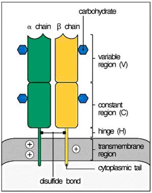
(Reproduced from Murphy K et al. (2008) Janeway’s Immunobiology (7th Ed.) Garland Science, New York.)
Structure of the γ−δ T-cell receptor
This is a second type of heterodimer linked by a disulphide bridge, expressed on a small subpopulation of T cells that make up less than 5% of all T cells (in peripheral blood, although they may be present in a higher proportion in secondary lymphoid tissues, mainly mucosal ones). It consists of extracellular immunoglobulin-type V and C regions, a hinge region, hydrophobic transmembrane segments and short cytoplasmic tails similar to those of the α-β chains. The hinge region contains cysteine residues involved in interchain disulphide bridges. The function of this type of receptor is still unclear, but it is less evolved and is known to recognise non-peptide molecules derived from bacteria as well as small phosphorylated molecules (phospholipids presented by CD1).
Lymphocytes with α-β receptors can express the co-receptor CD4 (Th) or CD8 (Tc), but never both simultaneously. T lymphocytes with a γ-δ receptor do not normally express a co-receptor and, if they do, it will always be CD8.
Constant proteins
The CD3 proteins and the ζ-ζ dimer associate non-covalently with the α-β or γ-δ heterodimers, and when the TCR recognises the antigen, these associated proteins transduce the signals that lead to T-cell activation.
CD3 proteins
The CD3 protein complex is made up of 3 types of protein: CD3-γ, CD3-δ and CD3-ε. These 3 proteins are organised to form two heterodimers alongside the TCR: CD3-ε/CD3-γ and CD3-ε/CD3-δ (Figure 7.3). The genes for these three proteins are located contiguously on chromosome 11, which suggests a common evolutionary origin. The N-terminal extracellular regions of the CD3-γ, -δ and -ε chains contain a single immunoglobulin-type domain, and for this reason all three belong to the immunoglobulin superfamily. The cytoplasmic domains of CD3-γ, -δ and -ε contain motifs with signalling functions called ITAMs (immunoreceptor tyrosine-based activation motifs).
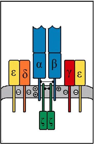
(Reproduced from Murphy K et al. (2008) Janeway’s Immunobiology (7th Ed.) Garland Science, New York.)
ζ-ζ dimer
TCR complexes also contain a homodimer of the ζ chain linked by a disulphide bridge. This ζ chain, although it is also transmembrane, has the greater proportion of the protein in the cell cytoplasm. The CD3 proteins and the ζ chain are identical in all T cells regardless of their specificity, which confirms that they are involved in signalling and not in antigen recognition.
The ζ chain has a short extracellular region, a transmembrane region and a long cytoplasmic region that contains three ITAMs. This chain belongs to the immunoglobulin superfamily.