Here is the syllabus explained in the video above. If you have any questions, post them in the comments section at the bottom of the page.
HLA class I antigens
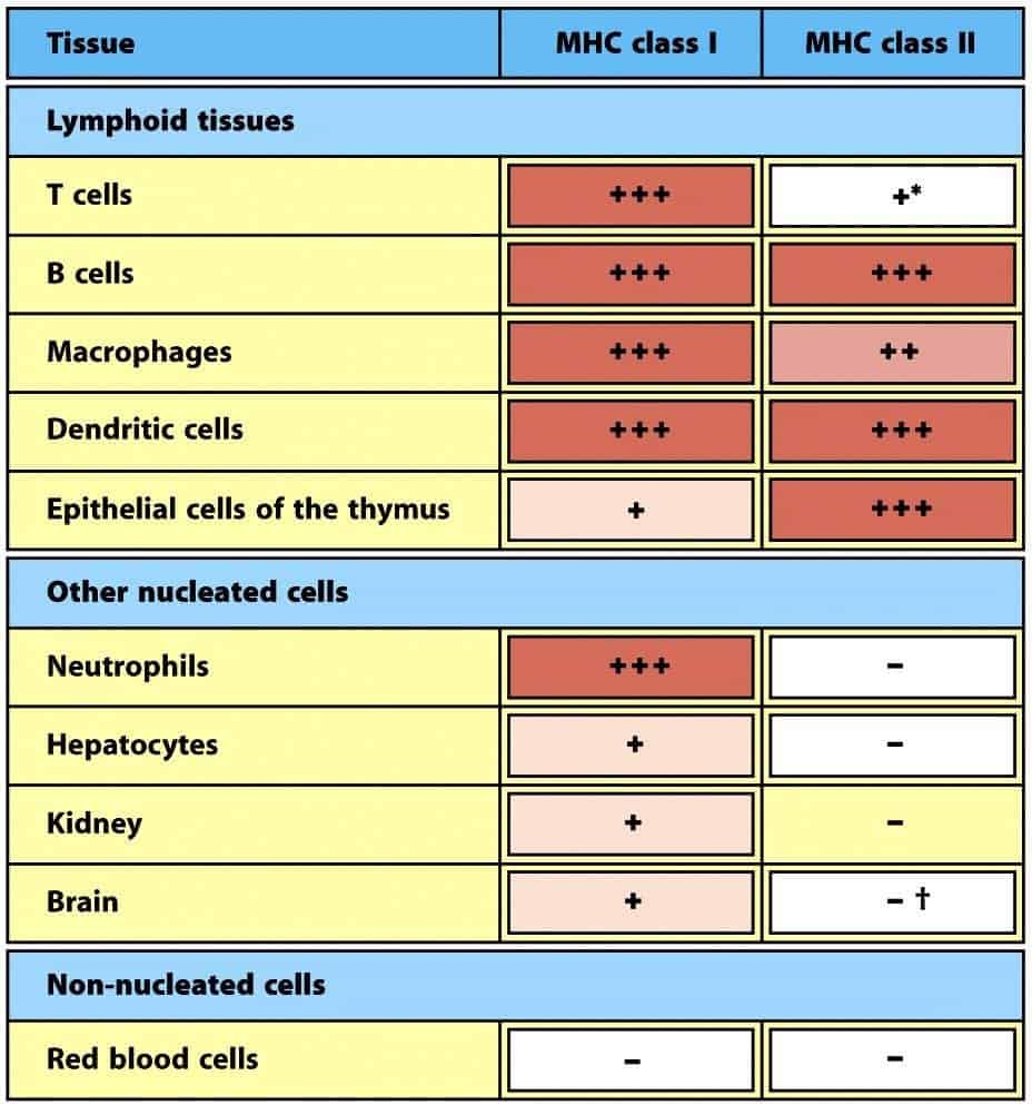
Human chromosome 6 carries multiple, highly polymorphic genes that encode the classical HLA class I antigens: HLA-A, -B and -C. The function of these three proteins is to present peptides to Tc cells, and their structure is very similar. HLA class I antigens are expressed on all cells of the body except erythrocytes (Figure 9.7). HLA I proteins are heterodimers made up of (Figure 9.3):
- A heavy or α chain, which is a transmembrane glycoprotein of about 45 kD and 300 amino acids. The alpha chain has 3 domains. The α1 and α2 domains, in the N-terminal portion, because of their particular folding, form a pocket or cavity (a physical gap) for peptide binding. The α3 domain is of the immunoglobulin type and is responsible for anchoring the HLA class I antigen to the plasma membrane.
- A soluble associated protein, non-covalently bound, called β2-microglobulin, with a single domain (of the immunoglobulin type) and encoded outside the HLA system.
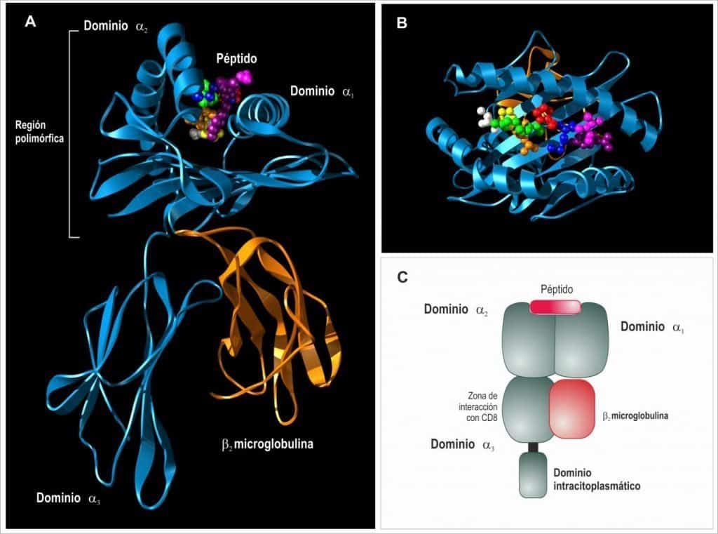
(A) Class I molecules are formed by a transmembrane chain called the α chain, non-covalently bound in its extracellular portion to a small protein called β2-microglobulin. (B) The α1 and α2 domains form a peptide-binding region called the peptide groove, which interacts with TCRαβ. (C) The α3 domain interacts with the CD8 coreceptor at the site indicated. (Reproduced from Regueiro J.R., López C., González S. & Martínez E.
(2011) Inmunología. Biología y Patología del Sistema Inmune. (4th Ed.) Editorial Médica Panamericana, Madrid.)
Peptide-binding cavity
The peptide-binding cavity is formed by the α1 and α2 domains, which build a flat base by folding into antiparallel β sheets, on which two α-helical structures rest.
For an HLA class I protein to be expressed on the cell surface, the peptide-binding cavity has to be filled by an antigenic peptide. If this pocket is not filled, the protein cannot be expressed. When these proteins were first studied, and were isolated, purified and crystallised to analyse their structure, it was thought that the crystals were not valid because they were always “contaminated” by a peptide. It was later found that this was not contamination, but that the peptide formed part of the complete structure of the protein.
HLA restriction
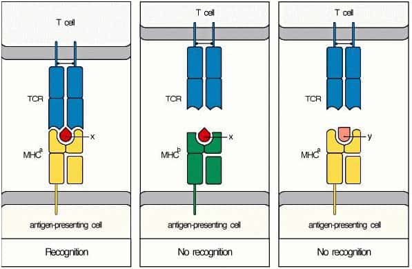
Antigen recognition by T cells is restricted by the HLA system. The specific antigen receptor of T cells (TCR, T-cell receptor) recognises the HLA + antigenic peptide complex. For the T cell to bind to the infected cell, therefore, the TCR must recognise both the presenting HLA protein and the peptide that it
holds in its pocket; if these two conditions are not met, binding does not occur. This combined recognition of the peptide and the HLA molecule is called HLA restriction, because the ability of the T cell to recognise the antigen is restricted. This explains the very high specificity of T cells.
(Reproduced from Murphy K et al. (2008) Janeway’s Immunobiology (7th Ed.) Garland Science, New York.)
HLA class II antigens
HLA class II antigens are heterodimers made up of two glycoproteins encoded by genes of the class II region of the MHC, located on the short arm of chromosome 6. The heavy α chain (34 kD) is encoded by the A genes, and the light β chain (29 kD) is encoded by the B genes (Figure 9.5). Both chains are transmembrane and associate through non-covalent bonds after their independent synthesis in the rough endoplasmic reticulum.
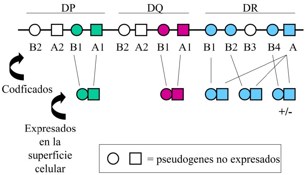
For each of the different HLA class II antigens (HLA-DP, HLA-DQ and HLA-DR) there is an A gene and a B gene. An exception to this rule is the HLA-DR proteins, whose system is more complex, with at least 3 HLA-DRB genes on the different chromosomes 6. Thus, multiple class II loci are expressed (up to 8 in humans) (Figure 9.6):
- DPA1, DPB1
- DQA1, DQB1
- DRA, DRB1, DRB2, DRB4
HLA class II antigens are expressed constitutively on professional antigen-presenting cells (APCs) and on thymic stromal cells (Figure 9.7).
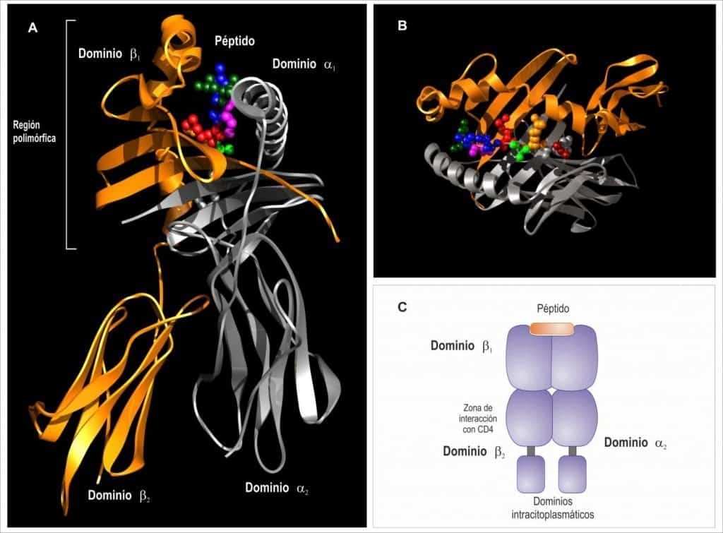
(A) Class II molecules are formed by two very similar transmembrane chains called the α and β chains. (B) The α1 and β1 domains form a peptide-binding region called the peptide groove, which interacts with TCRαβ. (C) The β2 domain interacts with the CD4 coreceptor at the site indicated. (Reproduced from Regueiro J.R., López C., González S. & Martínez E. (2011) Inmunología. Biología y Patología del Sistema Inmune. (4th Ed.) Editorial
Médica Panamericana, Madrid.)
(Reproduced from Murphy K et al. (2008) Janeway’s Immunobiology (7th Ed.) Garland Science, New York.)
In addition, they can be expressed inducibly on other cell types such as vascular endothelium or fibroblasts, under the action of certain signals such as IFN-γ (Figure 9.7). Their expression is therefore much more restricted than that of HLA-I antigens. Their function is to display exogenous antigens, which have followed the endocytic pathway, to helper T cells, activating them. These cells produce cytokines that act on various target cells.
Like HLA class I antigens, HLA class II antigens are highly polymorphic. The HLA system is therefore a complex genetic system with multiple loci and many different alleles at each. All the genes are highly polymorphic except HLA-DRA, which has a single allele. The following image shows the different genes for HLA class II antigens. Note that in some cases the genes are not functional (pseudogenes) (Figure 9.6). These properties (polygeny and high polymorphism) allow APCs to express on their surface a wide variety of HLA class II molecules, which will enable them to present a large number of different peptides to helper T cells.
Each of the chains of the class II antigens consists of two domains (Figure 9.5):
- The membrane-proximal domains (α2 and β2) have a characteristic fold and belong to the immunoglobulin superfamily.
- The membrane-distal domains (α1 and β1) build, through their folding, a physical gap for peptide binding.
The ribbon diagram shows that the α1 and β1 domains form a peptide-binding cavity or pocket in the same way as in HLA-I molecules, except that here this structure is formed by 2 different proteins joined non-covalently. To do so, these α1 and β1 domains fold into a flat region formed by antiparallel β sheets on which two α-helices rest (one from each protein). As in class I molecules, if this pocket or physical cavity is not filled with an antigenic peptide, HLA class II proteins cannot be expressed on the surface. The main difference from HLA class I antigens is that, in HLA class II molecules, the pocket is more flexible because it is formed by two proteins; in class I Ag it is a more rigid structure because it is formed by a single protein. This determines the type of peptide that can bind to each of the two molecules.