Here is the syllabus explained in the video above. If you have any questions, post them in the comments section at the bottom of the page.
Nomenclature of the HLA system
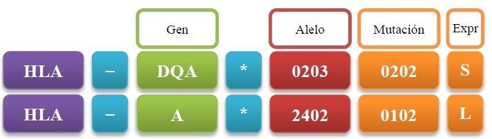
The antigens of the HLA system are named in a standardised way. The name begins with the letters HLA followed by a hyphen. Next, the gene in question is indicated by the letter that identifies it as class I (A, B, C, D, E, F, G) or class II (DP, DQ, DR). For class II, it must be specified whether the gene encodes the alpha chain (A) or the beta chain (B). Where there are several genes for that chain, as in the case of DRB, the gene number must be specified. Once the gene has been specified, an asterisk is written, followed by the allele of that gene present on the individual’s chromosome (Figure 9.8).
The allele is precisely identified by a system of at least four digits. The first and second digits indicate the allele group. The third and fourth digits indicate the specific allele within that group. The system can be supplemented with digits that provide more information: the fifth and sixth digits are used to indicate mutations in exons, and the seventh and eighth digits to indicate mutations in introns. Finally, a capital letter (L, N, Q, S) can be added, specifying the level of expression of the protein.
For example, the gene HLA-DRB3*0202 refers to the gene that encodes the type 3 beta chain of an HLA class II antigen of the DR type; specifically, the second allele of the second group of this gene.
Mechanisms of variability
A series of variability mechanisms make it extremely difficult to find two individuals in the population whose cells express the same collection of HLA antigens. These mechanisms are polygeny, codominance, polymorphism and complementation.
All these mechanisms make each individual’s combination of antigens a very reliable identifying mark, with a probability of error of the order of 1/106. Our cells present so many and such varied HLA antigens on their surface so that the greatest possible number of different antigenic peptides can be presented, ensuring that we have T cells with a suitable TCR. It should also be borne in mind that all these genes lie very close together, in a region of 1 cM of the short arm of chromosome 6. This means that the recombination rate is very low and, consequently, these genes tend to be inherited as a block, forming the so-called HLA haplotype. This, together with the great variability already explained, makes the analysis of HLA antigens very useful for various identification tests such as paternity tests.
Polygeny
Polygeny, or the presence of several different genes related to the same functions, ensures that each individual produces a wide variety of HLA molecules. For example, the HLA-A, B and C molecules have the same function and yet all of them are synthesised in each of our nucleated cells (Figure 9.9).
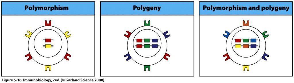
(Reproduced from Murphy K et al. (2008) Janeway’s Immunobiology (7th Ed.) Garland Science, New York.)
Codominance
The genes on both chromosomes are expressed. Thus both the HLA antigens inherited from the mother and those inherited from the father are found on the cell membrane (Figure 9.10). The consequence of polygeny and codominance is that any nucleated cell of the body can present up to six different HLA class I antigens: A, B and C inherited from the father; and A, B and C inherited from the mother.
Polymorphism
The genes that encode HLA antigens have a large number of different alleles. That is, there are many different forms of each of the genes (many forms of A, many forms of B and many forms of C for HLA class I) and each of us expresses two of these forms in our cells, one inherited from the mother and the other from the father.
The genes of the HLA system can be ranked according to their degree of polymorphism (Figure 9.11):
- HLA class I: HLA-B > HLA-A > HLA-C.
- HLA class II: HLA-DRB > HLA-DP > HLA-DQ. Among the HLA-DRB genes, the most polymorphic is HLA-DRB1.
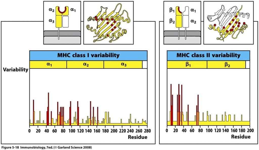
This variability is not distributed evenly along the whole gene but is concentrated in hypervariable regions, which coincide with the amino acids of the peptide-binding cavity (Figure 9.12).
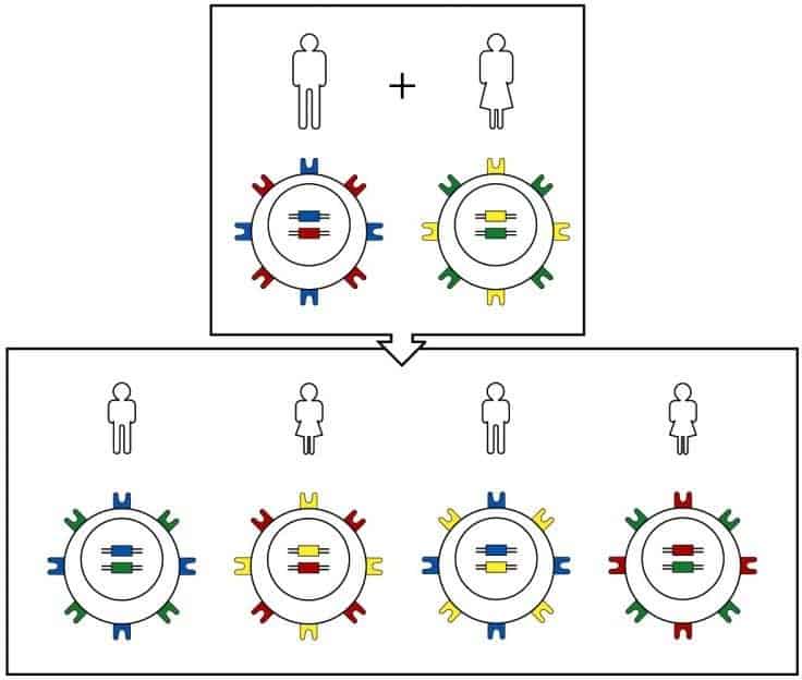
The MHC is so polymorphic that almost all individuals are likely to be heterozygous at each locus. In an individual, the alleles of both MHC haplotypes are expressed, and their expression results in the presence of their products on the cell surface. (Reproduced from Murphy
K et al. (2008) Janeway’s Immunobiology (7th Ed.) Garland Science, New York.)
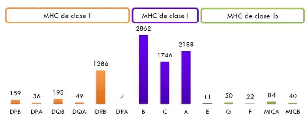
The graph shows the number of known alleles for each HLA locus. The most polymorphic loci are HLA-B and HLA-DRB. The numbers shown are merely indicative, since new alleles are constantly being discovered. The height of the bars represents the number of alleles of the HLA system assigned by the WHO Nomenclature Committee for Factors of the HLA System (updated January 2013).
(Reproduced from Murphy K et al. (2008) Janeway’s Immunobiology (7th Ed.) Garland Science, New York.)
Complementation
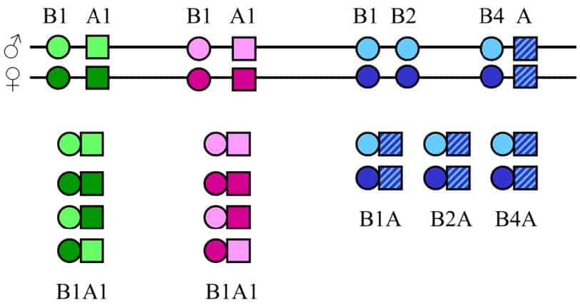
There is one last variability mechanism, present only in HLA class II antigens, called complementation (Figure 9.13). HLA class II molecules are composed of a beta heavy chain and an alpha heavy chain, which are synthesised independently in the endoplasmic reticulum. This means that the pairing of an alpha chain with a beta chain occurs at random, with two possible outcomes:
- Complementation in cis: a maternal alpha chain pairs with a maternal beta chain, or a paternal alpha chain pairs with a maternal beta chain.
- Complementation in trans: a maternal alpha chain pairs with a paternal beta chain, or a paternal alpha chain pairs with a maternal beta chain.
Physiology of the HLA system
The physiological function of HLA class I and HLA class II molecules is to present antigenic peptides, once the antigen has been processed, to Tc and Th cells respectively, since TCRs, unlike BCRs, are not able to recognise the antigen in its native state.
HLA molecules are also of great importance in other, non-physiological settings such as organ transplantation, in identifying individuals in forensic medicine, and in conferring susceptibility to certain autoimmune diseases. HLA molecules are only expressed on the cell surface when they are bound to the peptide, and are then called the antigenic complex.
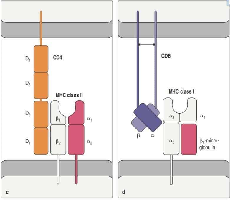
When an HLA class I molecule on the surface of any nucleated cell binds to its appropriate TCR, its peptide-binding cavity already contains the antigen peptide. The coreceptor of the Tc cell, CD8, associates with the α3 domain of the HLA class I molecule (Figure 9.14).
When an HLA class II molecule is expressed on the surface of an antigen-presenting cell, with the peptide already bound in its peptide-binding cavity, and binds to the TCR of the Th cell, the coreceptor, CD4, will bind to its natural ligand, which is the β2 domain of the HLA class II molecule (Figure 9.14).
(Reproduced from Murphy K et al. (2012) Janeway’s Immunobiology (8th Ed.) Garland Science, New York.)
The CD4 and CD8 coreceptors are glycoproteins that facilitate intercellular binding and also enhance signal transmission to the interior of the cell. Once the antigenic complex is recognised by the TCR, the T cells begin to produce cytokines, which act as chemical signals. The cytokines attract more T cells and induce B cells to proliferate and produce antibodies. The antibodies are released into the bloodstream to find and bind more antigens, so that pathogens cannot reproduce and cause disease.
(Reproduced from Regueiro J.R., López C., González S. & Martínez E. (2011) Inmunología. Biología y Patología del Sistema Inmune. (4th Ed.) Editorial Médica Panamericana, Madrid.)
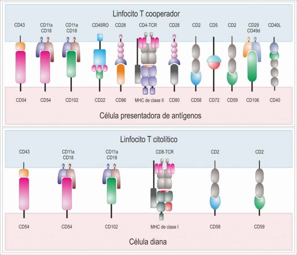
In many cases the binding of the antigenic complex to the TCR is not enough to produce an effective response, and the participation of a series of accessory molecules is required, whose function is to contribute to the development of an effective immune response by facilitating the interaction between the different cells. An immunological synapse is thus formed between T cells and the antigen-presenting cell, which is more intense between helper T cells and antigen-presenting cells than the one formed between cytotoxic T cells and their target cells (Figure 9.15).
A wide variety of molecules take part in the immunological synapse, notably:
- Specific antigen receptors: BCR and TCR.
- Co-receptors CD4 and CD8.
- Antigen-presenting molecules: HLA class I and class II.
- Receptors for adaptor molecules (opsonins): receptors for complement proteins (CD21) and Fc receptors for immunoglobulins (CD16).
- Cytokine receptors: for IL-1 (IL-1R or CD121a) and for IL-2 (CD25).
- Co-stimulatory molecules involved in activation and differentiation: CD28 (T cells) and CD80 or CD86 (on the APC), CD19, CD20 (B cells), CD2 (T and NK cells), CD45 (pan-leucocyte marker); CD40 (on the APC or B cell) and CD40L (on the T cell).
- Molecules induced after activation: CD25, CD38 and CD69.
- Cell adhesion molecules: CD2, CD11(a,b,c), CD18.
- Markers of the state of cell differentiation: CD45R0 (memory) or CD45RA (naive).