Redness, heat, swelling, pain and loss of function appear. All these signs reflect the function of the inflammatory reaction: attracting the cells and factors needed to fight the infection.
Celsus described inflammation as the combination of rubor, calor, tumor and dolor (redness, heat, swelling and pain). Later, Galen added another feature, loss of function, as a consequence of inflammation and pain.
- Rubor: redness.
- Calor: increased temperature in the area.
- Tumor: swelling.
- Dolor: activation of the nerve endings in the area.
Cells and mediators of acute local inflammation
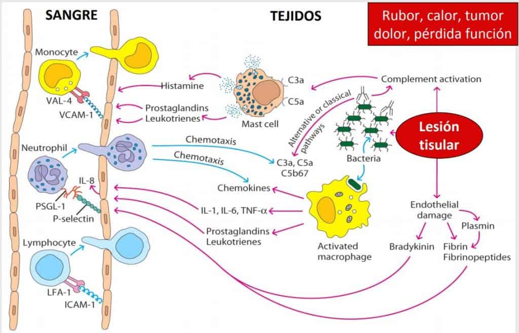
Tissue injury induces the formation of complement products (which act as opsonins, anaphylatoxins and chemotaxins). Macrophages phagocytose bacteria, become activated and release cytokines and other substances.
The initial tissue injury caused by trauma can be enough to set off an inflammatory response. When the skin is broken, microorganisms enter. In addition, the subcutaneous blood vessels are ruptured, that is, the endothelial cells of the broken vessels.
This tissue injury is able to activate complement, a group of proteins that circulate inside the blood vessels. These proteins are activated and the complement activation factors (anaphylatoxins: C3a, C5a) are formed, which have a direct effect on the endothelial cells of the blood vessels in the affected area: vasodilatation and increased permeability.
In addition, beneath the epidermis of the affected area there are phagocytes and mast cells that have receptors for the complement factors C3a and C5a. The receptors expressed on the mast cell membrane then react with the complement factors and release into the surrounding tissue a series of vasoactive substances such as histamine (which acts directly on the vascular endothelium), prostaglandins and leukotrienes.
Macrophages are also activated when microorganisms enter through the open wound, and they release various chemokines (IL-8) and three cytokines (IL-1, IL-6 and IFNα). These also have an effect on the endothelium.
All these substances act on the vascular wall, activating it and increasing the expression of adhesion molecules such as selectins and integrins. These substances are vasoactive and have a chemotactic effect, that is, they cause cells to extravasate and head for the area where the injury and infection by the microorganism have occurred. This results in swelling, or tumor, in the area.
It is very important to prevent microorganisms from reaching the blood, since this would cause SEPSIS, which has a vasodilator effect in many parts of the body, leading to death from hypovolaemia and multiple organ failure (the immune system prevents this).
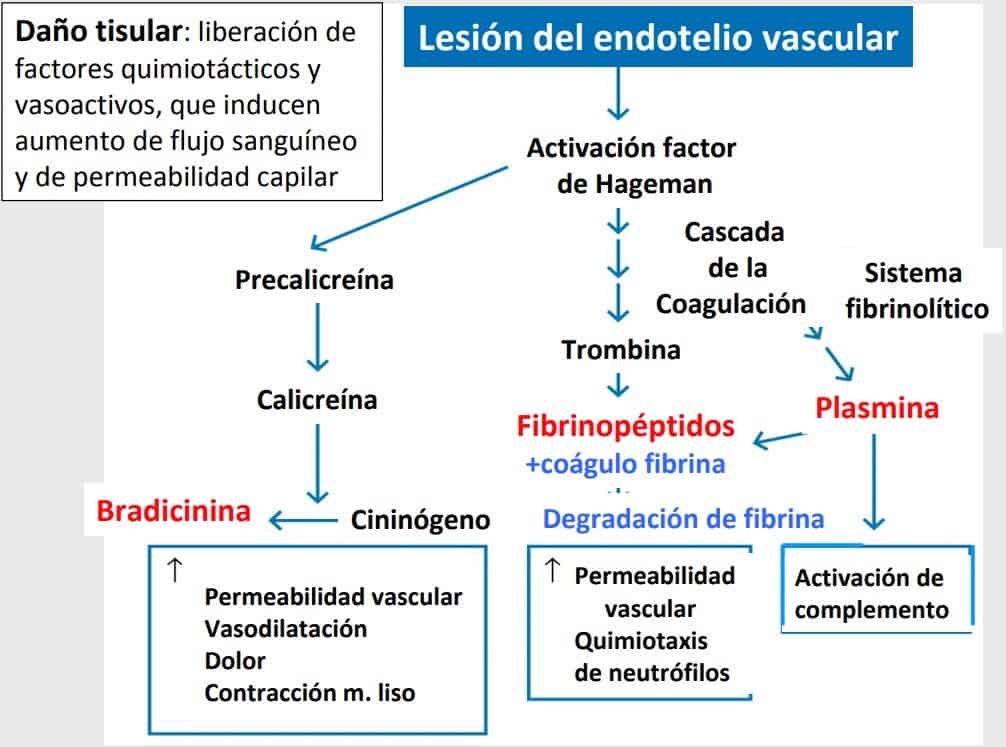
When the endothelial cells are broken, Hageman factor (factor XII) is activated, which in turn activates three other systems, similar to complement, that activate proinflammatory mediators:
- Kinin system: kinins contract smooth muscle and cause pain. The main component of the kinins is bradykinin, which has an important effect on vascular permeability, one of the decisive factors in allowing cells and fluids to reach the site of inflammation. (They constrict the passage of air in asthma by constricting the smooth muscle of the bronchial walls.) Bradykinin is responsible for the symptoms of local inflammation (redness, pain, etc.).
- Coagulation system: proteins (fibrinopeptides) are formed that give rise to blood clots, which break down to release more substances, then form another clot and break down again, and so on. This mechanism is due to changes in the vascular wall and is responsible for the intermittent bleeding that occurs when we injure ourselves.
- Fibrinolytic system: produces plasmin, which, by acting on the clot, releases elements that increase vascular permeability. Plasmin on its own is able to activate complement. In addition, plasmin acts on the fibrin clot and degrades it.
The common factor of these three systems is Hageman activation factor.
This whole system is maintained for as long as the microorganism remains in the individual. The defence system is not switched off until the causative pathogen has been destroyed. All these substances and cells generated at the site of infection produce pus, which consists of dead immune cells and pathological cells.
This process of pus formation also destroys nearby tissue, which leads to scarring. This is why this type of process (common in peripheral immunity) is not well suited to the intestine. An inflammatory process in the intestine would bring with it tissue destruction and loss of absorptive surface.
In short, these three systems (kinins, fibrinogen and coagulation), together with complement, are the four systems that generate proinflammatory mediators with specific effects.
In addition, tissue injury induces the formation of plasma mediators released by several interconnected systems: kinins, coagulation, fibrinolytic and complement. These mediators bring about local coagulation.
Vasodilatation occurs (producing swelling), cells are attracted from the circulation and blood flow slows down.
Chemotaxis reactions are produced by other factors released by macrophages (chemokines IL-1, IL-6 and TNF-α) and by complement.
The breakdown of the membrane phospholipids of various cell types (macrophages, monocytes, neutrophils, mast cells) gives rise to important mediators involved in the inflammatory reaction, such as the derivatives of arachidonic acid (thromboxane, prostaglandins, leukotrienes) and platelet-activating factor (PAF).
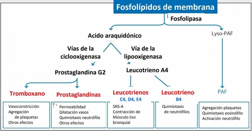
The release of phospholipids when the cell membrane of the cells of innate immunity (mast cells, macrophages, neutrophils, etc.) is broken will give rise, by two different pathways, to a whole series of substances (markers) that have been the therapeutic target of many anti-inflammatory drugs. These drugs are aimed at modifying the effects of many of these substances.
Thromboxanes and prostaglandins are formed through the cyclo-oxygenase pathway, and the 4 types of leukotrienes are formed through the lipoxygenase pathway. Platelet-activating factor (PAF) is also derived from membrane phospholipids.
The effects of both thromboxanes and prostaglandins are: increased permeability, coagulation and platelet aggregation, vasoconstriction secondary to the initial vasodilatation, and chemotaxis.
NOTE: leukotrienes, prostaglandins and thromboxane are derivatives of arachidonic acid.