Here is the syllabus covered in the previous video. If you have any questions, post them in the comments section at the bottom of the page.
Characteristics of antibodies
The minimal structure of an antigen that is recognised by an antibody and is capable of generating an immune response is called an epitope.
Epitopes may result from the sequence of amino acids in the primary structure (linear epitopes) or from the folding of the protein (conformational epitopes). Epitopes make close contact with the paratopes of immunoglobulins.
The three fundamental characteristics of an antibody are affinity, avidity and specificity (determined by the amino acid sequence of its variable domains).
Affinity determines how quickly the antibody (Ab) binds to the Ag and is determined by the strength of the bonds between the two molecules. This binding is never covalent and is in an equilibrium shifted to one side or the other according to the affinity. This binding strength, and therefore the affinity, depends on the number and type of chemical bonds between paratope and epitope.
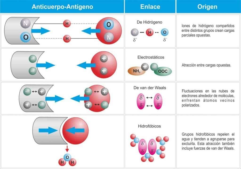
These bonds can be of several types (Figure 6.9):
- Hydrogen bonds: H ions shared by atoms of the Ag and Ab create opposite partial charges.
- Electrostatic bonds: charges of opposite polarity arise between Ab and Ag.
- Van der Waals forces: fluctuations in the electron clouds around molecules bring neighbouring polarised atoms face to face.
- Hydrophobic bonds: hydrophobic groups of the Ab and the Ag come together, excluding water molecules.
(Reproduced from Regueiro J.R., López C., González S. & Martínez E. (2011) Inmunología. Biología y Patología del Sistema Inmune. (4ª Ed.) Editorial Médica Panamericana, Madrid.)
Depending on the type and number of bonds, an antibody will have greater or lesser affinity for its antigen. Avidity is determined by the number of binding sites. It is the strength with which a multivalent antibody binds to a multivalent antigen. Some antigens have repetitive sequences in their structure. These antigens behave in a multivalent way, and the Ag-Ab binding strength is greater than the simple sum of the affinities of each of the antibody’s binding sites for the antigen. Thus, the greater the number of binding sites, the greater the avidity.
For example, pentameric IgM is usually the most avid for an antigen because it has 10 binding sites. In physiological situations, avidity is likely to be more important than affinity because antibodies are mostly divalent and antigens are most commonly multivalent. Specificity, avidity and affinity are determined by the Fab region of the antibody. Function is determined by the Fc fragment.
Functions of antibodies
Antibodies are good examples of how structure is closely related to function. They are Y-shaped molecules in which the short arms (Fab) interact with the antigen and the stem (Fc) provides the effector function. As a consequence of the Ab-Ag reaction, different functions will take place:
- Agglutination – neutralisation of the antigen. Antibodies are bivalent, that is, they have two antigen-recognition sites. This allows the antigen to be agglutinated, so that it can be eliminated more effectively. In addition, when the whole surface of the pathogen is coated with antibody molecules, a phenomenon called neutralisation occurs. For example, some toxins attack nerve endings, but when antibodies bind to them, they neutralise them.
- Opsonisation – phagocytosis. Coating of the pathogen surface by antibodies can have further consequences. Macrophages, which have receptors for the crystallisable portion of the antibody (Fc), called FcR, can phagocytose pathogens that have been coated with antibody. In this case the coating is known as opsonisation.
- Immobilisation of the pathogen. If the antibody binds to the motile part of the pathogen (cilia, flagella), it will immobilise it, reducing its pathogenicity.
- Complement activation. When an antibody binds to an antigen, a conformational change occurs in the Fc region of the antibody, which induces activation of the complement system. Only IgM and IgG are capable of this, playing an important role in adaptive immunity against infections. The recognition unit of the complement system that recognises bound antibodies is the C1q protein of the C1 factor.
- Expulsion. When the antibodies are of the IgE type and the antigens are parasites, IgE-antigen binding promotes the release of vasoactive amines that relax smooth muscle and cause diarrhoea in the intestine to expel the parasite.
- Antibody-mediated cytolysis (ADCC). Another function mediated by receptors for the Fc portion of the antibody is antibody-dependent cell-mediated cytotoxicity (ADCC). The effector cells are mostly NK lymphocytes, which, on recognising the antibody bound to the surface of the pathogen, release cytotoxic substances that attack the pathogen. The mechanism of ADCC is independent of the complement system and requires the NK lymphocyte to express FcR membrane receptors for the distal portion of IgG antibodies (CD16).
- Immunity in the fetus and neonate: during pregnancy and the first months of life, maternal immunoglobulins are the only specific defence mechanism the newborn has. Its immune system has not yet matured and it is initially unable to make its own immunoglobulins. These have to be transported across the placenta, or across the intestinal epithelium of the neonate, from breast milk.
Concentration and function of the different isotypes
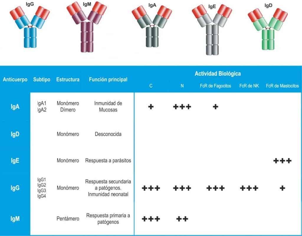
Each antibody isotype carries out some of these effector functions more effectively (Figure 6.10). Thus, with the same variable region, different constant regions are associated with differences in the effector function of the antibody:
- IgA: there are two subclasses, IgA1 (80-90% of the total serum IgA) and IgA2 (30-50% of the total IgA in secretions such as milk). The two subclasses appear to be regulated independently. Serum levels vary with age and are raised in women during lactation, as well as in chronic infections, liver disease and myeloma. A low IgA level indicates generalised protein deficiency or specific IgA immunodeficiency. This Ig is the most abundant in seromucous secretions such as saliva, milk or colostrum. It is part of the defence system of the external surface of the body. In secretions it occurs as a dimer joined to a J chain, which links the two IgA monomers, and to the secretory component, whereas in the blood it occurs as a monomer (made up of two alpha heavy chains and two light chains). Its main function is neutralising, although it also has, to a lesser extent, the capacity to activate complement.
- IgD: it is found mainly in plasma (less than 1% of the total plasma immunoglobulins, and undetectable in healthy individuals) and always as a monomer. Its function is unknown, but it is abundant on the surface of B lymphocytes and is thought to take part in the antigen-induced differentiation of these cells. B lymphocytes with IgD become an anergic cell (one that will never be activated, useless). It is also thought that it may be an evolutionary remnant that will disappear.
- IgE: its plasma concentration (in monomeric form) is low and it is undetectable in healthy individuals. It binds (through Fc receptors) to the surface of basophils and mast cells, and its function is defence against large parasites; however, in industrialised society it is associated with immediate hypersensitivity reactions such as asthma or hay fever (hygiene hypothesis).
- IgG: in the plasma or serum of normal adults, IgG accounts for 75% of the total circulating immunoglobulins. Serum IgG levels tend to vary with age, and even with race. A raised IgG level is associated with chronic liver disease and myeloma; reduced levels may be associated with malignancy and severe protein deficiencies, as well as with immunodeficiency. It has four subclasses, all of them monomeric. IgG1 is the most abundant in plasma, whereas IgG4 is the scarcest. It shows great functional versatility: it is important in fetal immunity because it crosses the placenta and enters the fetal plasma (the only antibody of the neonatal period and of maternal origin), in the secondary response and in complement activation, as well as enhancing phagocytosis, cytotoxicity and neutralisation.
- IgM: it is found in blood and in infected tissues in pentameric form (joined by J chains). Each unit consists of two µ heavy chains and two light chains. It is the most important isotype in the primary response. It is highly efficient against multi-determinant antigens such as bacteria and viruses, and acts in complement activation. It is a good neutralising antibody, although not as good as IgG and IgM. Raised IgM levels are associated with hepatitis, myeloma, Waldenström’s macroglobulinaemia, infections and hyper-IgM syndrome (a congenital immunodeficiency). Reduced IgM levels are seen in some antibody immunodeficiencies.
Therefore, in serum we will find, from most to least abundant: IgG, IgA, IgM; the amounts of IgD and IgE will be undetectable in a healthy individual (less than 1% of the total).
(Reproduced from Regueiro J.R., López C., González S. & Martínez E. (2011) Inmunología. Biología y Patología del Sistema Inmune. (4ª Ed.) Editorial Médica Panamericana, Madrid.)
Transport of IgA to mucosal surfaces
Immunoglobulin A is the main isotype in secretions, especially in the epithelia of the digestive and respiratory tracts. IgA-producing plasma cells (the final stage of B-lymphocyte differentiation) are found predominantly in the connective tissue (lamina propria) immediately beneath many epithelial surfaces.
IgA synthesised in the lamina propria is secreted as dimeric IgA associated with a joining chain, the J chain (Figure 6.11). This polymeric form of IgA binds selectively to a polyimmunoglobulin receptor (poly-Ig-R) present on the basolateral surfaces of epithelial cells. Once dimeric IgA has bound to this receptor, the complex is internalised into the cell and transported through the cytoplasm of the epithelial cell in transport vesicles to the apical part of the cell. This process is called transcytosis.
Once at the apical surface of the epithelial cell, the poly-Ig receptor is cleaved by an endoprotease (favoured by the acidic pH of the intestinal lumen), releasing the outermost portion of the receptor still bound to the dimeric IgA. This receptor fragment released together with the IgA is called the secretory component, and it appears to protect dimeric IgA from possible enzymatic degradation; the other fragment remains attached to the surface of the epithelial cell.
The tissues with the greatest IgA synthesis are the intestine, the respiratory epithelium, the breast (during lactation) and other exocrine glands such as the salivary and lacrimal glands. The main function of IgA antibodies is thought to be to protect the epithelial surface from infectious agents by neutralising them.
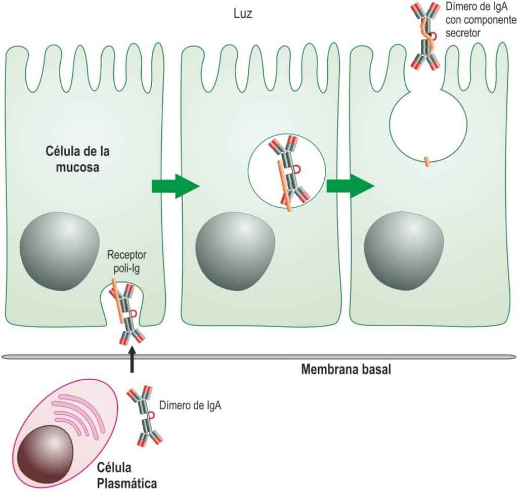
Figure 6.11 Transport of IgA to mucosal surfaces
(Reproduced from Regueiro J.R., López C., González S. & Martínez E. (2011) Inmunología. Biología y Patología del Sistema Inmune. (4ª Ed.) Editorial Médica Panamericana, Madrid.)