Here is the syllabus covered in the previous video. If you have any questions, post them in the comments section at the bottom of the page.
Immunoglobulin isotypes
Small variations in the amino acid sequence of the constant regions of the light and heavy chains (which affect the size, charge and solubility of the protein) define different subtypes of the chains (isotypes):
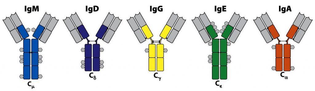
- 5 types of heavy chain are known: μ, δ, γ, α and ε. They differ from one another in structure and function.
- There are also 2 types of light chain: κ and λ. They are structurally and functionally equivalent.
Igs take their name from the heavy chain, regardless of the type of light chain they carry. Thus we have IgM, IgD, IgG, IgA and IgE, respectively (Figure 6.5).
In addition, small variations within the molecules of the IgG and IgA isotypes make it possible to distinguish four subclasses of IgG (IgG1, IgG2, IgG3 and IgG4; with γ1, γ2, γ3 and γ4 heavy chains, respectively) and two subclasses of IgA (IgA1 and IgA2; α1 and α2 chains). Overall, therefore, there are nine Ig isotypes, which may contain κ or λ light chains.

(Reproduced from Murphy K., Travers P. & Walport M. (2008) Janeway’s Immunobiology (7th Ed.) Garland Science, New York.)
(Reproduced from Regueiro J.R., López C., González S. & Martínez E. (2011) Inmunología. Biología y Patología del Sistema Inmune. (4ª Ed.) Editorial Médica Panamericana, Madrid.)
Differences between the Ig classes (Figure 6.5):
- IgE and IgM molecules have a greatly reduced hinge region and have a fourth constant domain (CH4).
- Each Ig also has glycosylation sites, represented by hexagons. IgA and IgD are the most heavily glycosylated; IgE and IgM have intermediate glycosylation, and IgG is the least glycosylated (Figure 6.5).
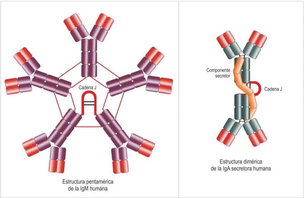
- Two classes can appear in multimeric form (Figure 6.6), forming additional structures. Thus IgM (in serum) usually occurs as pentamers (5 basic structural units), which together add up to 10 heavy chains and 10 light chains. The five Igs are joined by their Fc regions with a J chain (J = joining), leaving the antigen-binding sites free. In total it can recognise ten independent identical Ags. IgA usually occurs as dimers joined by a J chain different from that of IgM. IgA can also appear with another associated peptide (the secretory component, which gives it resistance to acid) to form secretory IgA (sIg), which plays an important role at mucosal surfaces. In total it recognises four independent identical Ags on the surface of pathogens.
Igs can also show different allotypes (minor polymorphic or allelic variations in the amino acid sequences of the constant regions of the heavy and light chains, which segregate according to Mendelian genetics).
Variability of immunoglobulins
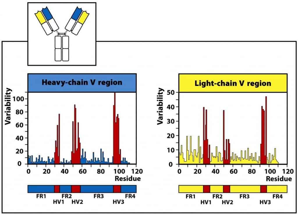
The variable domains are those that specifically recognise the antigen. In each variable domain the protein folds so as to leave three points of contact with the antigen. These points are called hypervariable regions (HV) or CDRs (complementarity-determining regions). The less variable regions that flank them are called framework regions (FR) (Figure 6.7).
(Reproduced from Murphy K., Travers P. & Walport M. (2008) Janeway’s Immunobiology (7th Ed.) Garland Science, New York.)
Some of the amino acids in the hypervariable regions CDR1, CDR2 and CDR3 can show 100% variability from one Ig to another. The area of close contact with the antigen (the junction of the CDR portions of the heavy and light chains) is called the paratope, which pairs with the epitope of a given antigen. Each of these CDR segments is 5-10 amino acids long. The extreme variability found in the V regions of immunoglobulins reflects the need to recognise a large number of antigenic structures. Most of the contact points correspond to the third hypervariable region (CDR3), where the greatest variability accumulates.
Genetic organisation of immunoglobulins
Each Ig chain (heavy or light) is encoded by a set of genes. The variable domain is encoded by V, D and J genes (in the heavy chains) or only V and J genes (in the light chains). The constant domains are encoded by a single gene (C). There is one C gene for each of the heavy-chain isotypes (Figure 6.8), each of which is denoted by a “C” followed by the Greek equivalent of the class:
- IgM – Cμ, there is only 1 gene for this chain.
- IgD – Cδ, there is 1 gene for this chain.
- IgG – Cγ, there are 4 genes: γ1, γ2, γ3 and γ4.
- IgE – Cε, there is 1 gene for this chain.
- IgA – Cα, there are 2 genes: α1 and α2.
Resting B cells transcribe RNA that includes the first 2 constant genes (Cμ and Cδ). Through alternative RNA processing, the cells can simultaneously process RNAs for IgM and IgD, both with the same antigen specificity, since they contain the same VDJ rearrangement.

(Reproduced from Regueiro J.R., López C., González S. & Martínez E. (2011) Inmunología. Biología y Patología del Sistema Inmune. (4ª Ed.) Editorial Médica Panamericana, Madrid.)