Here is the syllabus covered in the previous video. If you have any questions, post them in the comments section at the bottom of the page.
The specific immune response is based on the specific recognition of antigens, both in B-cell and in T-cell responses. Each type of lymphocyte has fundamental proteins for this specific recognition, and immunoglobulins are the antigen-specific molecules of B cells. The main function of the B lymphocyte is therefore the synthesis of immunoglobulins.
Immunoglobulins are a large group of proteins found in serum and other body fluids. They exist in two main forms:
- A soluble form, which is then called antibodies.
- Anchored in the membrane of B lymphocytes, forming part of the B-cell antigen receptor (BCR).
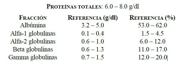
Immunoglobulins are found in the gamma fraction of serum proteins after they are separated by electrophoresis on a protein electrophoretogram (Figure 6.1).
Immunoglobulins
Basic protein structure
Immunoglobulins share a common structure of 4 polypeptide chains, identical in pairs. This structure consists of two heavy chains (H = heavy) and two light chains (L = light), identical in each immunoglobulin molecule (Figure 6.2). The molecular weight of the H chains ranges from 55 to 77 kDa and that of the L chains is 25 kDa. The different chains are stabilised by disulphide bridges between the two heavy chains and between each heavy chain and a light chain (but never between two light chains).
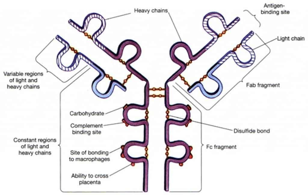
Both heavy and light chains have a basic structural unit (the immunoglobulin domain), which is repeated 4-5 times in the heavy chains and 2 times in the light chains. This domain consists of two beta sheets, each made up of 3-4 antiparallel alpha helices, which are very stable thanks to hydrophobic interactions and an intradomain disulphide bridge between two cysteines, each belonging to one of the helices of each sheet. Proteins with this motif in their structure are said to belong to the so-called immunoglobulin superfamily.
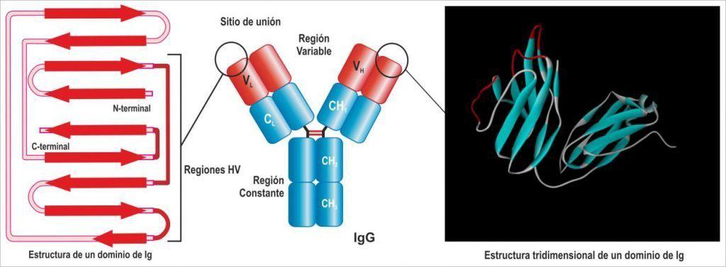
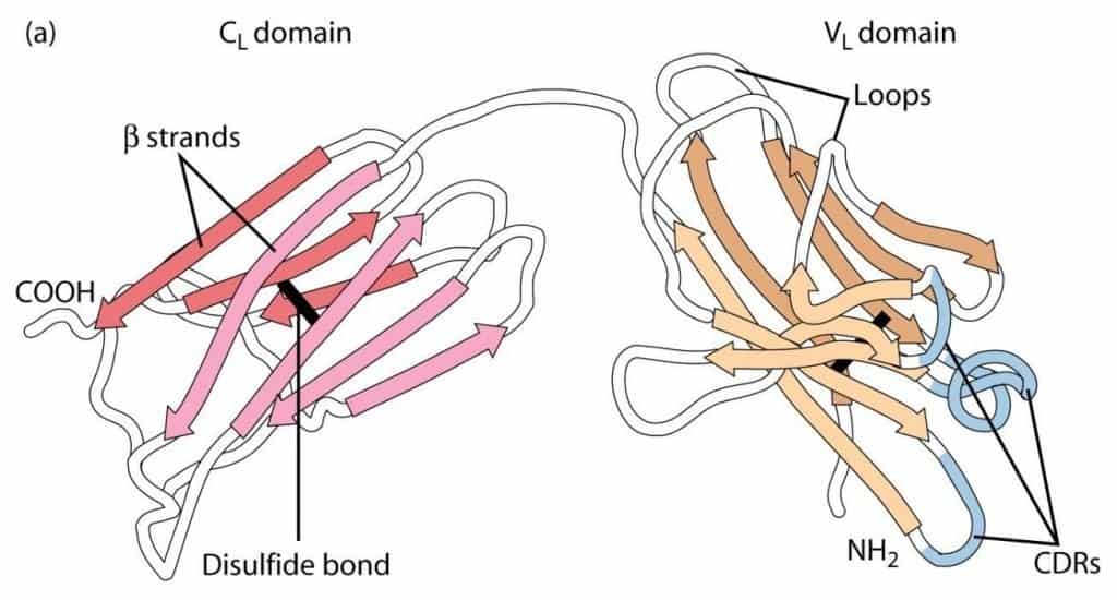
(Reproduced from Regueiro J.R., López C., González S. & Martínez E. (2011) Inmunología. Biología y Patología del Sistema Inmune. (4ª Ed.) Editorial Médica Panamericana, Madrid.)
These domains are named according to whether or not they come into contact with the antigen (Figure 6.3). The domains in the carboxyl-terminal region are called constant (C) domains, and a subscript (H or L) indicates whether they belong to the heavy or the light chain: hence the CH and CL domains. The amino-terminal domains are responsible for antigen recognition and are the variable (V) domains; again, a subscript indicates whether they belong to the heavy or the light chain: VH and VL. Only in the heavy chains (since the light chains have a single constant domain) are the constant domains numbered: CH1, CH2, CH3. There may be an additional constant domain (CH4), as in IgM and IgE. The H chain is therefore made up of 4-5 domains: the first, at the amino-terminal end, is variable and the following 3 or 4 are constant. Light chains always consist of 2 domains: one variable and one constant.
Between the first and second constant domains of the heavy chain there is a hinge region of variable length (10-60 amino acids), not organised into domains, where the disulphide bonds between the heavy chains form and which gives the Ig flexibility. This is what gives the molecule its characteristic structure, abbreviated as a Y. Each Ig can recognise 2 identical Ags simultaneously, because each pair of variable domains (VH and VL) interacts with an Ag identical to that of the other pair.
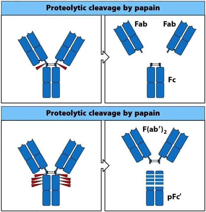
When the Ig molecule is subjected to the action of plant proteases (pepsin and papain), different protein fragments are released (Figure 6.4):
- Papain: a cut is made in each heavy chain, at the level of the hinge region. This gives 3 fragments: one containing the 2-3 constant domains of the 2 heavy chains, called the crystallisable fragment (Fc), and 2 identical fragments that include the variable domains of both chains, one constant domain of the heavy chain and the whole of the light chain, called Fab (antigen-binding fragment). The specificity of the Ig resides in Fab, and Fc determines the function of the molecule.
- Pepsin: cuts are made at several points in the heavy chains. This gives one large fragment with the two antigen-binding sites joined together, called F’(ab)2, and several fragments of the constant portion of the heavy chains, called pFc’.
(Reproduced from Murphy K., Travers P. & Walport M. (2008) Janeway’s Immunobiology (7th Ed.) Garland Science, New York.)