For the immune system to work properly, it must distinguish between self and non-self. There is a regulatory mechanism that has been conserved throughout evolution: apoptosis. The neuroendocrine system is involved in this regulation.
To control the discrimination between self and non-self there is a mechanism called tolerance. This phenomenon allows the immune system to recognise, but not mount immune responses against, the body’s own antigens.
Most of the lymphocytes that reach the thymus, the bone marrow or the germinal centres of the lymph nodes die. Approximately 95% of these cells do not proliferate, in order to eliminate autoreactive cells.
A distinction is made between central tolerance, which takes place in the primary lymphoid organs (bone marrow and thymus, affecting T and B cells), and peripheral tolerance, outside the bone marrow and thymus.
Central tolerance
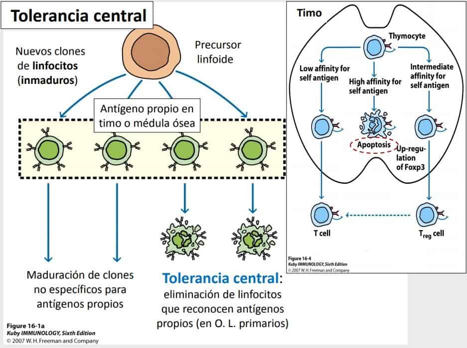
This is the elimination of lymphocytes that recognise self antigens and takes place in the primary lymphoid organs.
T cells leave the thymus when they are immunocompetent, that is, when they have rearranged competent receptors capable of recognising foreign antigens in the context of the body’s own HLA molecules. This takes place in the thymus, where two selection steps occur:
- In positive selection, those lymphocytes with useful receptors survive, capable of recognising the MHC antigens on the membrane of thymic cells. The cells of the thymic epithelium express AIRE proteins + self antigens, which must be recognised by the immature thymocytes. If they do not recognise them, it is not worthwhile for these cells to mature. That is, there must be a certain degree of affinity for MHC molecules.
- In negative selection, lymphocytes that recognise self Ag with high affinity undergo apoptosis within the thymus (clonal deletion).
The mechanism of cell activation mediated by apoptosis for thymocytes that recognise self antigens is not well understood.
The strength with which the lymphocyte binds to the self Ag presented by MHC molecules is thought to be important. Weak binding of the lymphocyte to the self Ag leads to the elimination of the lymphocyte, because it needs to recognise self antigens.
High affinity leads to apoptosis of the lymphocyte, as it would be potentially autoreactive. Only those that recognise the self antigen with intermediate affinity will mature, and leave as naive lymphocytes for the secondary lymphoid organs. This ensures that lymphocytes that would cause some kind of autoimmune disease (which have been increasing lately) do not leave the thymus. Thus, only 5% of thymocytes enter the circulation as mature lymphocytes.
Something similar happens in the lymph nodes and the bone marrow. B cells that undergo somatic hypermutation are cells that improve their affinity for the antigen, and only those with the highest affinity can compete for the antigen presented by follicular cells and are the ones selected.
Those cells that recognise antigens with the appropriate affinity survive.
Peripheral tolerance

In the periphery, control of the activation of T or B cells is also necessary, because the primary lymphoid organs, where lymphocytes acquire immunocompetence, do not contain all the possible self antigens of the body against which lymphocytes need to develop tolerance. That is, lymphocytes may encounter antigens in peripheral tissues that they had not seen before during their maturation, because they are found only in certain tissues.
It is an antigen-induced tolerance mediated by a series of cells. Cells acquire this property in a particular tissue depending on local factors in the tissue, which determine whether they become regulatory T cells. These mechanisms aim to ensure that in the periphery only foreign antigens are recognised, above all antigens capable of replication (pathogenic microorganisms).
These peripheral mechanisms are responsible for maintaining tolerance and are:
- Apoptosis of all those cells with specificity for self antigens. This is the most effective mechanism because it destroys the cell but does not cause inflammation.
- Anergy occurs when a lymphocyte that recognises an antigen presented by MHC class II molecules is activated without the appropriate costimulatory molecules to control the activation of that cell. That is, in the absence of costimulation, the lymphocyte becomes anergic (functional inactivation). For example, when we introduce APCs from another individual, even if they present the antigen specific for those T cells, they will not cause their activation. The specific T cell recognises specific foreign antigen but always in the context of the body’s own MHC molecules, that is, presented by a cell from the T cell’s own organism. When that does not happen, the process of anergy is triggered, or functional inactivation of those cells, which does not necessarily lead to their destruction.
**Although many cells die in the thymus all the time, when its tissue is examined it is clean. This is because macrophages and phagocytes reach it and clear and destroy the cell debris derived from cells lysed by apoptosis.