All the cells of the body have mechanisms for setting apoptosis in motion. It is a “supposedly” dangerous system and therefore has a safety mechanism that consists of keeping the proteases or factors that trigger apoptosis latent, not active (caspases are the proteins involved in all apoptotic processes).
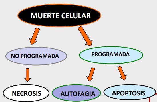
There are other, non-programmed processes that lead to cell death:
- Necrosis: this is the premature accidental death of cells in living tissues. It is irreversible. It is caused by the release of cell fragments that activate inflammatory processes. It can result from interruption of the blood supply.
Programmed cell death can occur through:
- Autophagy: the cell itself releases the contents of its lysosomes (proteases, lytic enzymes) into the cytoplasm, causing self-degradation. The cell digests itself. It is a mechanism for saving energy and obtaining building blocks for tissue. A distinction is made between microautophagy and macroautophagy.
- Apoptosis: a genetically controlled cellular process by which cells induce their own death in response to certain stimuli. It differs from necrosis in that apoptosis does not involve inflammation of the tissue. That is: IT DOES NOT BRING ABOUT AN INFLAMMATORY RESPONSE. The cells die and collapse, forming apoptotic bodies.
*Apoptosis is not exclusive to humans and is mediated by caspases.
Caspases
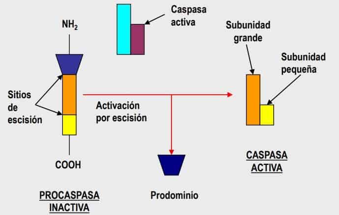
These are proteases that take part in apoptosis: they cause protein degradation until apoptotic bodies form, which are subsequently phagocytosed. Inside cells they are found as zymogens or pro-caspases (inactive form), which need proteolytic cleavage to become active. This proteolysis gives rise to the active form of the caspases (caspase 3), which is ultimately responsible for tissue destruction.
They have a defined structure:
- N-terminal domain:
- Long: initiator caspase.
- Short: activator caspase
- Catalytic region: C-terminal region.
Factors that trigger their activation: they are activated by proteolysis (proteases) and act in a cascade.
- Initiator caspases: they set the process in motion.
- Effector caspases: those that determine the formation of apoptotic bodies.
Apoptosis pathways
It can be activated by two kinds of signal:
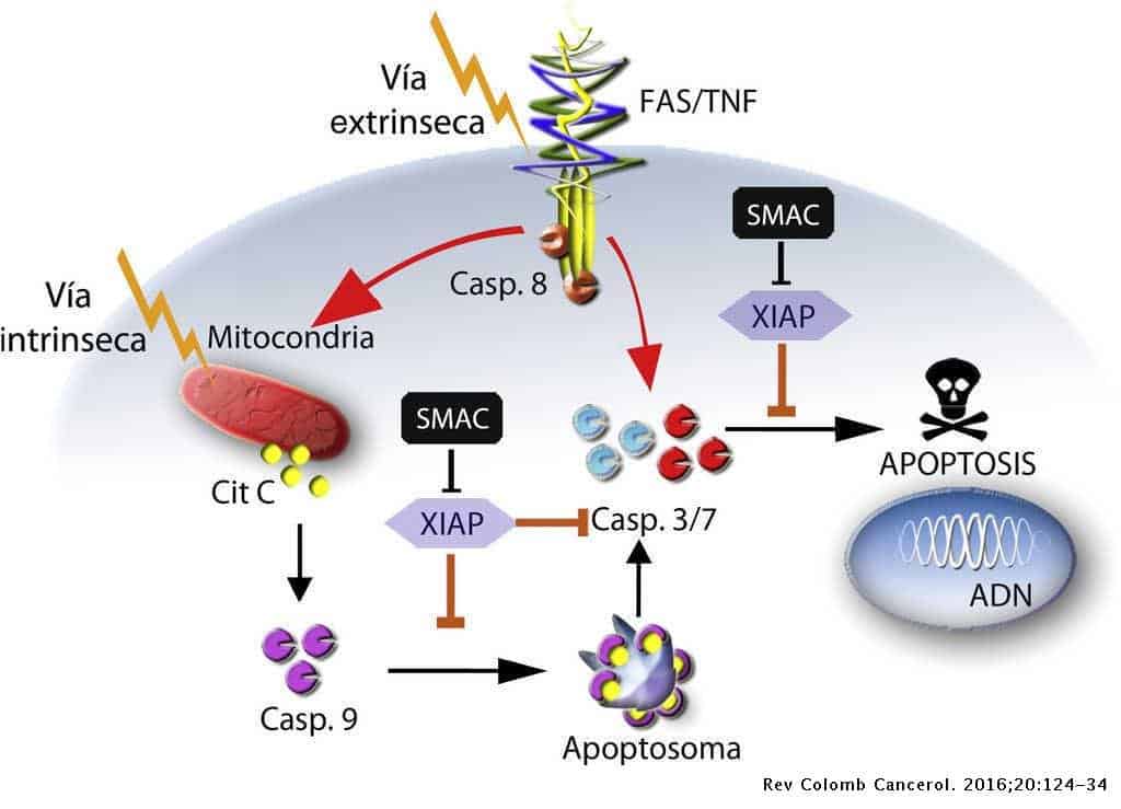
- Extrinsic: This is the most important one. It involves the binding of ligands to receptors, as in the case of Fas or TNF. These are molecules related to apoptosis. Cytolytic CD8+ cells and NK cells can also exert their function by secreting granzymes (an effect similar to Fas, Fas ligand: a hole in the cell destroys it, causing apoptosis). There are other signals related to integrins.
- Intrinsic: There are many signals that can lead to cell stress and thus to apoptosis. Radiation and UV rays, chemotherapeutic agents, viral infections, heat, osmotic changes, high concentrations of calcium, deprivation of growth factors (which can cause a cell to stop growing and enter apoptosis), oxidative stress and the formation of free radicals (ageing).
These two pathways can converge through caspase 9.
Apoptosis is activated by 2 pathways: the caspase is initially large but is broken down by a process of proteolytic cleavage to form a smaller active structure that sequentially activates more molecules, giving rise to a cascade.
Extrinsic pathway
takes place outside the cell, predominantly in the periphery. It covers everything that acts on the cell membrane.
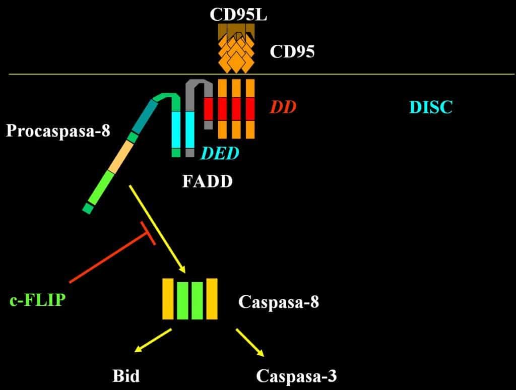
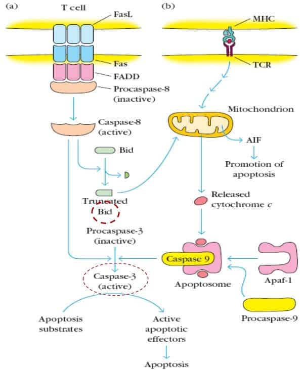
- It begins with the expression of FasL (Fas ligand) by a T cell, which is expressed persistently in any location in the body. FasL binds to Fas on the membrane of another cell and induces the trimerisation (fusion) of Fas, forming a Fas trimer.
- Recruitment of DISC (death-inducing signalling complex) then takes place; it is found in the cytoplasm and binds to the cytoplasmic domain of Fas. This interaction causes the expression and binding of FADD, an adaptor molecule responsible for attaching a pro-caspase 8 to the Fas trimer. This results in the activation (proteolysis) of pro-caspase 8, which becomes caspase 8, and this in turn will activate caspases 3, 6 and 7 (the final effector caspases). This complex would therefore be an initiator caspase.
- The extrinsic pathway also interacts with the intrinsic pathway. Caspase 8 is able to activate proteins of the Bcl-2 family (Bid protein), which causes the release of cytochrome C and Apaf-1 from the mitochondria, which will form a structure called the apoptosome (intrinsic pathway) that will set in motion an independent process of apoptosis.
Intrinsic (mitochondrial) pathway
This is a direct pathway that is set in motion in situations of stress or particular situations of the mitochondria, which release a series of proteins into the surrounding medium.
- The proteins of the Bcl-2 family control the formation of a pore in the mitochondrial membrane and the release of cytochrome C from the membrane.
- Cytochrome C is a determining factor in apoptosis by the intracellular pathway. It binds to two proteins found in the cell cytoplasm:
- Apaf1 (apoptotic protease-activating factor)
- Pro-caspase 9, which is activated to caspase 9.
- This binding gives rise to a structure called the apoptosome.
- The apoptosome (cytochrome C + Apaf-1 + caspase 9) cleaves pro-caspase 3 into caspase 3 (which activates effector caspases), triggering cell apoptosis.
Activation of caspase 9 acts as the link between the two apoptosis pathways.

In this context, what happens with T cells? (Description of the image)
T cells could undergo processes that affect both pathways.
A T cell that expresses Fas ligand (for example, a cytolytic lymphocyte) binds to a cell that expresses Fas on its membrane. This extrinsic mechanism is the one that would be set in motion in the case of cytolytic cells. They cause dimerisation of Fas, binding of FasDD, activation of pro-caspase 8, and caspase 8 is able to cause apoptosis per se. But activation of caspase 8 also causes the release of proteins of the BCL family that act on the mitochondria, releasing cytochrome C (and so on up to caspase 9).
The intrinsic pathway per se is the one that is set in motion especially in the thymus. We have the MHC class II molecule. Immature lymphocytes that express an appropriate TCR on their membrane will bind to APCs within the thymus and, depending on the strength of the interaction between the MHC class II molecule and the ability to recognise self antigens, these cells will die. They probably die when the strength of recognition is very high. This means that this process (MHC interaction with TCR) would lead to the release of cytochrome C from the mitochondria (binding to Apaf, caspase 9 and finally apoptosis). However, there is a gap in our knowledge of how this activation of the intrinsic pathway occurs.
Apoptosis in the immune system
In which situations is apoptosis relevant?
- Elimination of autoreactive B cells in the bone marrow and in peripheral tissues.
- Formation of the repertoire of non-autoreactive T cells in the thymus.
- T cells whose TCR does not recognise the body’s own HLA molecules die by apoptosis.
- T cells whose TCR recognises the body’s own HLA molecules with high affinity die by apoptosis.
- Somatic hypermutation: after it takes place in the germinal centres, B cells that fail to produce better Ab than the original die by apoptosis. Those that produce Ab of higher affinity differentiate into plasma cells (to synthesise Ig) or into memory cells.
- Cytolytic mechanism: induced by Tc lymphocytes (CD8+).
- Tolerance: lack of response to some foreign Ag (from the diet…). Specific lymphocytes are eliminated by apoptosis (and regulatory T cells).
- Homeostasis: after an infection has been cleared, the effector cells die so that the immune system returns to homeostasis. T cells are activated and Fas is expressed on the membrane.