This is a property common to all mucosal tissues. Cells enter the blood, extravasate and recirculate for several days; after this time they return to the mucosa, to the effector sites (epithelium or lamina propria).
Precursors of IgA-producing cells, activated T cells and APCs carrying antigen leave via the efferent lymphatics to the mesenteric lymph nodes and, through the thoracic duct, reach the bloodstream, where they recirculate and extravasate into the intestinal mucosa and four other exocrine tissues.
If we have a lymphoblast that has been induced, there are three molecules on its membrane that allow it to be identified. The process is regulated by the selective expression on lymphocytes of the integrin α4β7 and the chemokine receptor CCR9, which interact with the vascular addressin MAdCAM-1 and CCL25, expressed on vascular endothelial cells and on intestinal enterocytes respectively, but not in other lymphoid tissues. On activation they also express LFA-1 (the receptor for ICAM). The memory cell expresses only α4β7 and LFA-1.
A subpopulation of T cells express the integrin αEβ7 (CD103) and are retained in the epithelium by interacting with E-cadherin.
At the effector sites (LP), terminal differentiation into IgA-producing plasma cells is favoured by CD4+ T cells that produce TGF-β and other cytokines (IL-5, IL-6, IL-10), and by factors released by local APCs, with an important role for retinoic acid. The T cells produce TGF-β which makes the intestine behave in such a way as to generate tolerance-type responses.

Antigen can also enter through the epithelium, of the villi. Whatever the route of entry, it will always encounter a series of antigen-presenting cells.
Enterocytes are not professional APCs under normal conditions, but some epithelia can be activated and begin to express MHC class II antigens. The antigen-presenting cells migrate to the mesenteric lymph nodes, where the immune response is generated in a more organised and specific way.

This migration also occurs in other mucosal structures of MALT. In the nasal mucosa (NALT), for example, microorganisms cross the epithelium and meet APCs, which activate lymphocytes, and these leave into the circulation through the thoracic duct. Once they are recirculating, they will return to areas close to the focus of infection, the place where they were activated, since this is the most likely place for an infection to occur again.
| In the lymph circulation system there is a point of passage between the blood and the lymph, where already activated and differentiated cells leave into the blood and recirculate for about two weeks. But how do they know where they have to return to? The functional state of the dendritic cell, with its costimulatory signals and cytokines, induces the formation of adhesion molecules characteristic of the area where the cells were activated. An effector/memory lymphocyte expresses LFA-1 and α4β7, which is characteristic of activated cells of the intestinal mucosa. There are vascular addressins expressed on the endothelium of the vessels and on the endothelium of the intestinal mucosa, MAdCAM-1 and ICAM-1 and 2. Ig is produced in the healthy individual, above all IgA. In addition, B cells reach not only the lamina propria but also the intestinal epithelium. CCR9 is able to reach as far as the epithelium, the contact zones in the lumen of the intestine. |
Populations of the effector sites
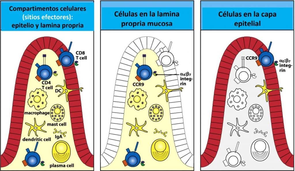
The effector sites (epithelium and mucosal lamina propria) contain different types of cells.
- Lamina propria: the main subtype of T cells is CD4+ + TCRαβ, cytokine-producing cells (responsible for inducing Th1 and Th2 responses), IgA-producing plasma cells (in some cases IgM), and also dendritic cells, macrophages, eosinophils, neutrophils…
- Epithelium: the main subtype of T cells is CD8+ + TCRαβ/TCRγδ, CD3 cells (known as IEL cells → intraepithelial lymphocytes). There are also NK cells and Paneth cells. APCs are less numerous. These cells are mainly responsible for monitoring the state of epithelial cells; if the cells are infected or are tumour cells, they destroy them so that the epithelium is renewed. A high percentage of CD8 cells are αβ, but naturally there is also a high number of cells with a γδ receptor, related to some diseases such as coeliac disease. These γδ cells are also watching what happens in the surroundings, looking for molecules that indicate cellular stress (MICA and MICB).
Most responses are of the Th2 type.
The lamina propria is prepared to produce inflammation if an infection occurs. However, an inflammatory reaction would be harmful in the intestine, since mucosa, and therefore absorptive area, can be lost. This situation of inflammation occurs, for example, in Crohn’s disease.
To avoid an inflammatory reaction, there are numerous anti-inflammatory signals in the lamina propria, since the function of absorption predominates over that of defence. Hence the importance of distinguishing between the antigens generated by food and those that genuinely have pathogenic capacity.
It processes information and changes its conformation (from latent to activated) in response to danger signals. An infection may be triggered, or a tolerogenic response may occur.