Effects of cytokines
They regulate cell activity in a coordinated and interactive way:
• PLEIOTROPY: one cytokine has several different effects on different target cells (e.g. IL-2 has several effects on different target cells, depending on the tissue in which it is found and on the receptor cells)
• REDUNDANCY: two or more cytokines have similar effects, which may vary from one tissue to another. Others have the same effects but are different cytokines. It is difficult to assign an effect to a single cytokine (e.g. chemokines that act in flocks)
• ADDITIVE EFFECT: the effect of two cytokines together equals the sum of the effects of each. (multiplier effect: that is, not just the sum of the effect of the two)
• SYNERGY: the joint effect exceeds the additive effect of the two cytokines separately; that is, one cytokine together with another will have a certain effect, different from that of the original cytokine with a different one.
• ANTAGONISM: one cytokine will have an inhibitory effect on the other.
The only two inhibitory cytokines: IL-10 AND TGF-β (transforming growth factor β)
- IL-10 acts by blocking antigen-presenting cells, and therefore blocking T cells
- TGF-β: acts by directly blocking the T cell, preventing it from being activated or proliferating
They are the only ones that act to produce a net inhibitory effect. However, there are other cytokines that normally act as positive cytokines and nevertheless block a second cytokine, acting as antagonists.
• CASCADE INDUCTION: some important cytokines induce a pyramid effect, a cascade of secretion of different cytokines. (one cytokine acts on a cell, which in turn induces another cytokine, and so on progressively; very typical of inflammatory responses)
In this way, the action of a cytokine on its target cell causes the latter to secrete one or more cytokines which, acting in turn on other target cells, induce the secretion of further cytokines.
- Two examples of this “cascade induction” property involve tumour necrosis factor alpha (TNF-α) and interleukin 1 (IL-1), which, when produced systemically, cause loss of homeostasis and lead to septic shock.
The medical interest here lies in manipulating the immune system, manipulating all of these cytokines and making the immune system block their function in certain situations, such as autoimmune pathologies, which occur when the immune response is activated in inappropriate tissues. These cytokines are very useful in this type of situation.

Image taken from the slides provided by Professor Arranz, Faculty of Medicine, Valladolid, Topic 13, Immunology, academic year 2014-15
Regulation of the effects of cytokines
There are 3 main factors that regulate the action of cytokines during the immune response, and they ensure that the effects do not have non-specific repercussions:
1.- The expression of receptors on target cells occurs only after activation by the antigen. A cell that does not express receptors cannot recognise cytokines. In the case of cells, sometimes the more immature they are, the less able they are to interact with other cells because they express fewer receptors on their surface; therefore, this expression determines the specificity of the cytokine.
2.- In some cases, a direct interaction between the cytokine-producing cell and its target cell is required. This ensures that the effective concentration gradient occurs only in the local microenvironment.
The paracrine effect is the most common: for example, a dendritic cell or macrophage presents an Ag to the T cell and releases IL-12, which will help this T cell to develop and mature towards a particular functional pathway. This paracrine effect, therefore, rests on this concept: it depends on the microenvironment and the concentrations of that cytokine in that medium, or on the degree of affinity of the receptor for the cytokine
3.- The half-life of these molecules in body fluids is usually very short; they act and, to avoid unwanted effects, that protein is destroyed. If not all pathogens are destroyed in an immune response, the immune response remains active and so does the cytokine; the half-life is therefore a factor controlling the immune response, its intensity and its duration. Cytokines last as long as the immune response is active. CLASSIFICATION OF CYTOKINES (PATTERNS) CLASSIFICATION OF CYTOKINES (PATTERNS)
Classification of cytokines (patterns)
1. Cytokines of the innate immune response
Murphy K., Travers P. & Walport M. (2008) Janeway’s Immunobiology (7th Ed.) Garland Science, New York.
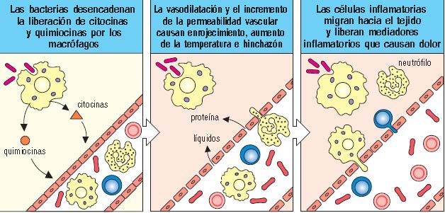
-INFLAMMATORY OR MACROPHAGE CYTOKINES: TNF-α (tumour necrosis factor alpha), IL-1, IL-6, IL-12: Macrophages produce cytokines as part of their function of warning other defensive cells of the presence of pathogens. The cytokines and chemokines released by activated macrophages start the process known as inflammation. When macrophages encounter bacteria or other types of microorganism in the tissues, they release cytokines that increase the permeability of blood vessels, allowing fluid and proteins to pass into the tissues. They also produce chemokines, which direct the migration of neutrophils towards the site of infection. The thickness of the endothelial cells of the blood vessel wall also changes, so that cells adhere to the wall and are able to move slowly along it. The accumulation of fluid and cells at the site of infection causes the redness, swelling, heat and pain that are collectively known as inflammation.

Murphy K., Travers P. & Walport M. (2008) Janeway’s Immunobiology (7th Ed.) Garland Science, New York
These three cytokines (the first three) are among the most pro-inflammatory in all processes. All three are produced by pathogens with the aim of attracting more cells to the inflammatory focus.
IL-12: (remember that IL-10 has an inhibitory effect on T cells by blocking antigen-presenting cells, which in turn blocks the synthesis of IL-12. It activates T cells so that they continue to produce certain cytokines that are relevant in responses to intracellular parasites.
-ANTIVIRAL INTERFERONS (TYPE I): IFN-α, IFN-β: all cells of the body, when infected by a virus, release type I interferons, which interfere with the ability of viruses to produce their own proteins using the cell’s machinery once it is infected. (Ebola virus survives because it blocks interferon synthesis)

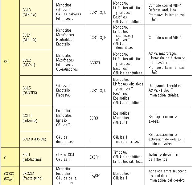
2. CHEMOKINES (chemotactic cytokines): Chemokines function mainly as chemoattractants for leucocytes; they recruit monocytes, neutrophils and other effector cells from the blood to sites of infection. They can be released by many different cell types, and serve to guide the cells involved in innate immunity to sites of infection. They also guide lymphocytes in adaptive immunity. They are heavily involved in inflammatory processes where containment or destruction is needed to finish off the invaders.
Chemokines are a family of proteins with similar structures that bind to chemokine receptors, which form part of an extensive family of G protein-coupled receptors.
Chemokines can be produced by a wide variety of cell types in response to bacterial products, viruses and agents that cause physical damage, which sets in motion the production of chemokine gradients that can direct phagocytes to the sites where they are needed.
CCL3: MACROPHAGE INHIBITORY PROTEIN (CC protein ligand, and when R: receptor)
Chemokines are classified mainly into two related but distinct groups. The CXC chemokines, whose genes are found mainly in a cluster on chromosome 17 in humans, have one amino acid residue (X) between two invariant cysteines (C) in the amino-terminal region. The CC chemokines, which in humans are encoded for the most part in a region of chromosome 4, have these two invariant cysteines adjacent to each other.
All the chemokines that attract neutrophils have this motif, whereas almost all the other CXC chemokines, even those that interact with CXCR3, 4 and 5, lack it. receptors and affects one or more cell types.
Murphy K., Travers P. & Walport M. (2008) Janeway’s Immunobiology (7th Ed.) Garland Science, New York.

Murphy K., Travers P. & Walport M. (2008) Janeway’s Immunobiology (7th Ed.) Garland Science, New York.
3. CYTOKINES OF THE SPECIFIC IMMUNE RESPONSE Th0, Th1 (IL-2, IFN-γ, IL-12), Th2 (IL-4, IL-5, IL-10, IL-13. CD4 cells activate the release of cytokines (they are the orchestra conductors) and, depending on the type of invading microorganism, the T cell is activated and differentiates to synthesise a particular cytokine profile:
- If, for example, we have an extracellular bacterium entering the blood, the Abs could act against it.
However, an intracellular parasite that enters a macrophage requires detection and activation by CD4+ cells, which in turn activate other cells capable of forming phagocytes (e.g. macrophage).
This explains why there are different subtypes (Th1, Th2…). These cells, also called cytotoxic, act by recognising the viral proteins of a cell invaded by a virus and bring about its destruction.
Th0: an immunocompetent but naive, non-activated cell. When it reaches a lymph node and recognises, for example, the Ag of an intracellular bacterium (inside a macrophage), it releases IL-12, which will cause T cells to differentiate into a type of cell that is more efficient against intracellular infections.
If B cells are activated: IL-4 and production of Th2. These Th2 cells are also important in allergic individuals (IgE)
-IL-12: causes T cells to differentiate into a specific subtype to eradicate that type of microorganism
-Th17: important above all in acute inflammatory responses.

Image taken from the slides provided by Professor Arranz, Faculty of Medicine, Valladolid, Topic 13, Immunology, academic year 2014-15.
4. HAEMATOPOIETIC GROWTH FACTORS SCF, IL-3, IL-7, GM-CSF (granulocyte-macrophage colony-stimulating factor), IL-11 (thrombopoietin), Epo (when erythrocytes increase, the capacity to oxygenate the muscle is much greater… cyclists)
SCF: they bind to bone marrow cells that do not yet have a defined fate, so that they only help these cells to be maintained.
These are cytokines that stimulate the production of haematopoietic cells.
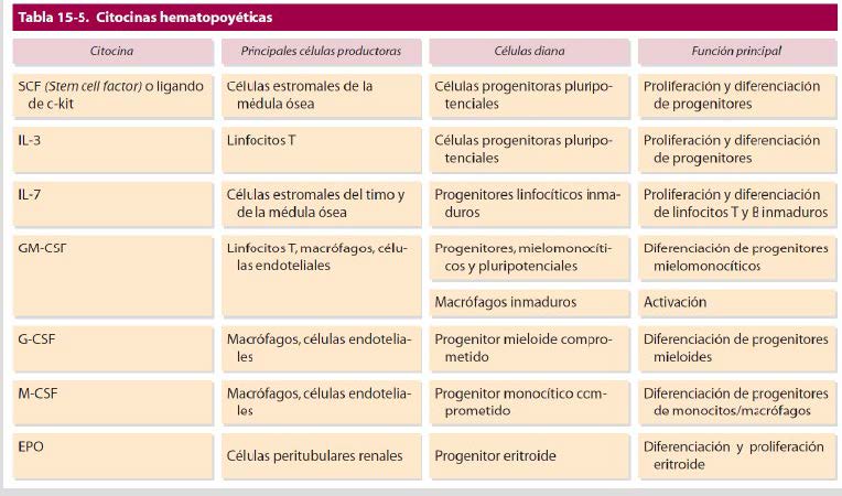
Image taken from the slides provided by Professor Arranz, Faculty of Medicine, Valladolid, Topic 13, Immunology, academic year 2014-15.
The cytokine network
Let us suppose we are in a secondary lymphoid organ: depending on the microorganism detected and the cytokines generated, these cells will go on to differentiate (Th1…2…17…); what cytokines do is promote the activity of all the cells of the immune system and others outside it (lymphoblasts) so that they take part in the inflammatory response.
GENERAL MODEL: The cytokine is recognised by the receptor (in general, membrane receptors are dimers) and, on binding, dimerisation occurs, which leads to the activation of a cascade of kinases that also activates, and causes dimerisation of, a family of STAT proteins, which act in the nucleus on DNA, activating a specific set of genes
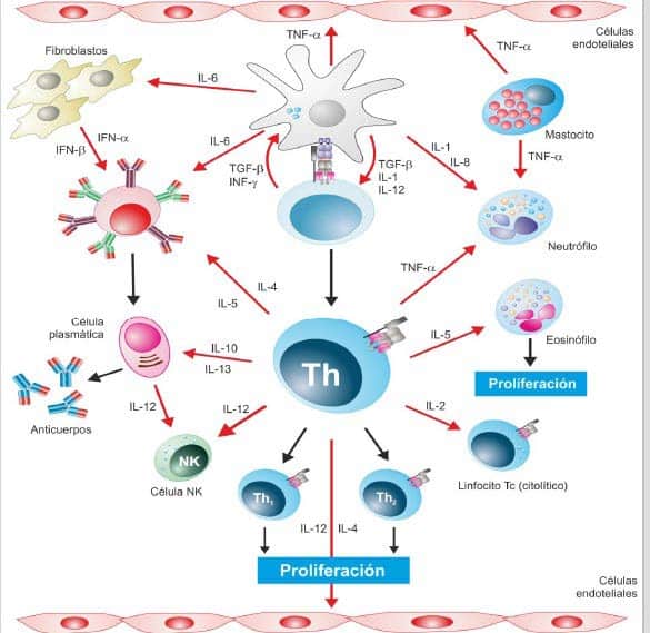
Image taken from the slides provided by Professor Arranz, Faculty of Medicine, Valladolid, Topic 13, Immunology, academic year 2014-15.
Each member of a family is a variant with a different specificity, which performs a particular function in the cell that expresses it:
- The haematopoietin receptor superfamily includes homodimeric and heterodimeric receptors, which are subdivided into families on the basis of protein sequence and subunit structure.
- The receptor for the macrophage cytokine IL-6 (chap. 2) is also a member of the haematopoietin superfamily, but it signals through an accessory chain other than βc or γc.
- The receptors for interferons and interferon-like cytokines are heterodimeric receptors that make up another, smaller family of cytokine receptors.
- The tumour necrosis factor receptor (TNFR) and the chemokine receptor family, the latter belonging to the very large superfamily of G protein-coupled receptors.
Many members of the IL-2 receptor subfamily (receptors for class I cytokines) share a signal-transducing γ chain (common gamma chain). The IL-2 and IL-15 receptors are heterotrimers in which the transducing chains are β and γ.