Here is the outline explained in the previous video. If you have any questions, post them in the comments section at the bottom of the page.
Preferences in peptide binding to MHC molecules
MHC class I molecules
MHC class I molecules have a peptide-binding groove formed by a floor of antiparallel β sheets on which two α helices rest (Figure 10.4-AB). Because this groove is formed by the α1 and α2 domains of the MHC class I α chain, that is, by a single protein, it is a very rigid site for peptide binding.
In addition to this limitation, peptides must meet other requirements in order to bind to MHC class I:
- Length: peptides that bind to MHC class I range from 8 to 11 amino acids in length, those of 9 amino acids being preferred.
- Ends: peptides bind to MHC class I molecules through their amino-terminal and carboxy-terminal ends.
- Critical anchor residues: MHC class I molecules contain sub-pockets that determine which types of amino acid can bind to them (aromatic, hydrophobic, non-polar, etc.). In this way, MHC class I molecules determine which peptides can bind (Figure 10.4-CD, Figure 10.5, Figure 10.6-CDE).
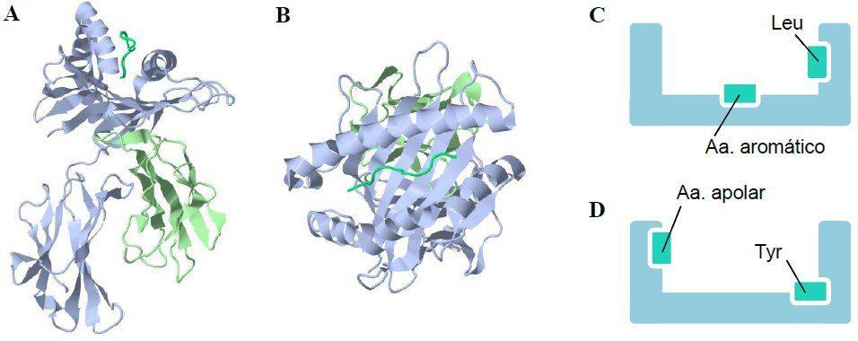
(A) (B) Ribbon diagram of MHC class I, side view (A) and top view (B). In both cases, an antigenic peptide can be seen in the peptide-binding groove. (C) (D) Diagram of the peptide-binding groove of MHC class I molecules showing the critical anchor residues of the antigenic peptides. (C) represents the molecule in Figure 10.5-A. (D) represents the molecule in Figure 10.5-B. (Courtesy of David Pérez Torres, University of Valladolid.)
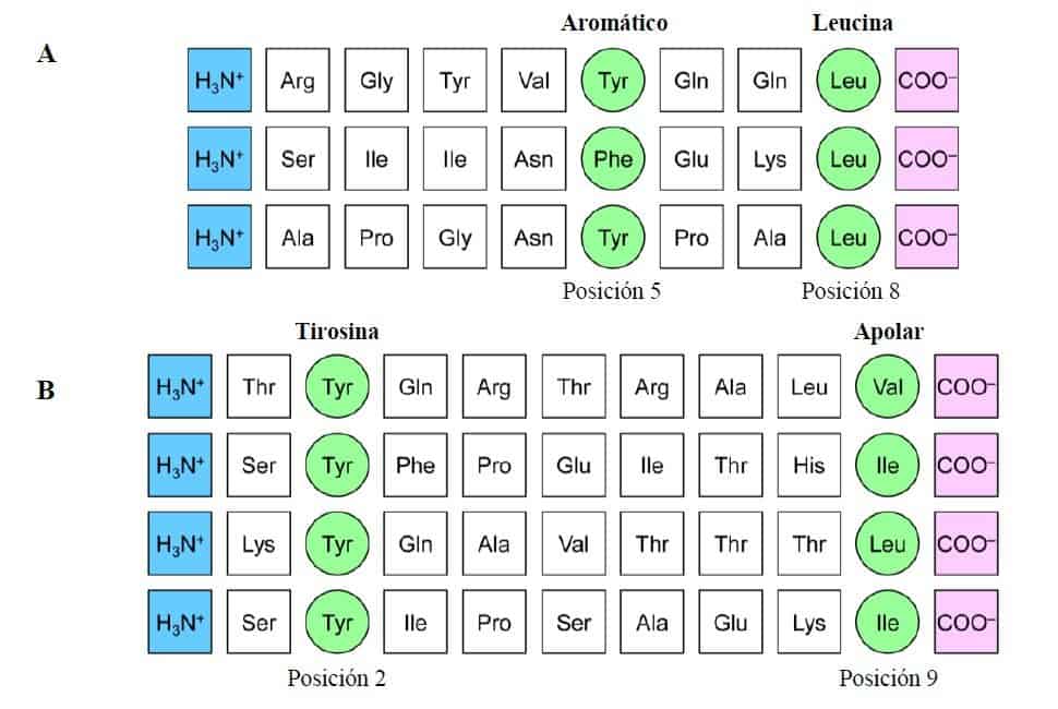
The figure shows the amino acid sequences of peptides eluted from two different MHC class I molecules. The anchor residues (shown in green) differ for peptides that bind to different alleles of MHC class I molecules, but are similar for all peptides that anchor to the same molecule. The peptides also bind to the amino-terminal (shown in blue) and carboxy-terminal (shown in pink) ends of MHC class I molecules. (A) Peptides that bind to this MHC class I molecule do so through anchor residues that need not be identical but are related: at position 5 they are always aromatic and at position 8 always leucine. (B) In this other MHC class I molecule, the bound peptide must contain tyrosine at position 2 and a non-polar amino acid
at position 9. (Redrawn and modified from Murphy K et al. (2008) Janeway’s Immunobiology (7th Ed.) Garland Science, New York.)
MHC class II molecules
The peptide-binding groove of MHC class II molecules is composed of two different molecules joined non-covalently, the α and β chains, which makes it a more flexible groove than that of MHC class I molecules (Figure 10.6-AB). This allows the peptides that bind to be up to more than 20 amino acids long, so they frequently hang over both sides of the groove. The peptide normally binds to MHC class II through 9 amino acid residues. As with MHC class I, there are critical anchor residues (Figure 10.6-CD
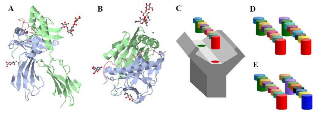
(A) (B) Ribbon diagram of MHC class II, side view (A) and top view (B). In both cases, an antigenic peptide can be seen in the peptide-binding groove, together with the anchoring position of other molecules. (C) Schematic representation of the binding of a nonapeptide to an MHC class I or II molecule. Note that the critical anchor residues (shown in green and red) are different in each MHC molecule. (D) Schematic representation of a peptide with the same anchor residues as the one in Figure 10.6-C. (E) Schematic representation of a peptide with different anchor residues from the one in Figure 10.6-C, so that it cannot
bind to the MHC molecule in that figure. (Courtesy of David Pérez Torres, University of Valladolid.)
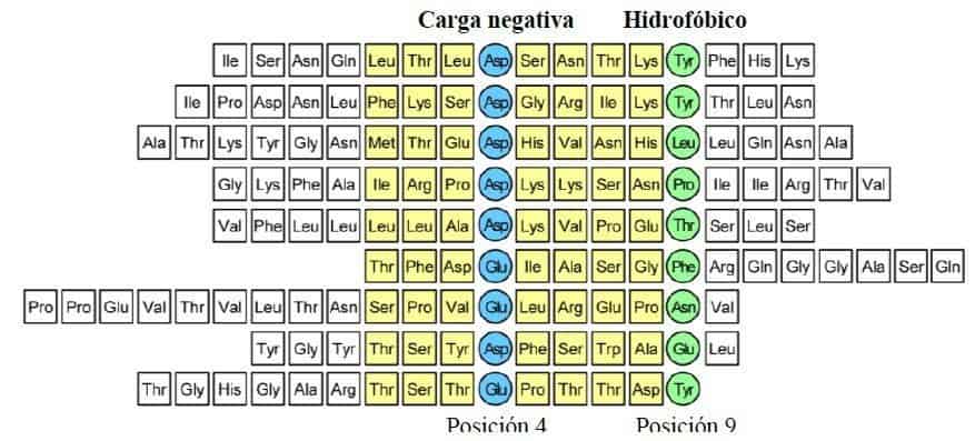
Sequence of peptides that bind to the HLA-DR3 allele of human MHC class II. The length of the peptides is variable and, by convention, the first anchor residue is numbered 1. The contact zone between the peptide and MHC class II comprises 9 amino acids. All the peptides share a negatively charged amino acid at position 4 (aspartic acid or glutamic acid) (shown in blue) and tend to have a hydrophobic residue at position 9 (shown in green). (Redrawn and modified from Murphy K et al. (2008) Janeway’s Immunobiology
(7th Ed.) Garland Science, New York.)
Superantigens
Conventional antigens are processed by antigen-presenting cells and presented to T cells on MHC class II molecules (Figure 10.8-A). In this case, TH cells with receptors able to recognise the antigen are activated and begin to produce cytokines. Activated TH cells stimulate only those B cells that react with the antigen against which the response is being generated, so that the immune system produces antibodies only against the antigen that was presented on the MHC class II molecule.
Superantigens (SAgs), such as Staphylococcus enterotoxin B (SEB), which causes food poisoning in humans, cannot be processed by antigen-presenting cells. Instead, they bind directly to constant regions of the MHC class II molecules of antigen-presenting cells and to constant regions of the TCR of TH cells, forming a bridge between the two molecular structures.
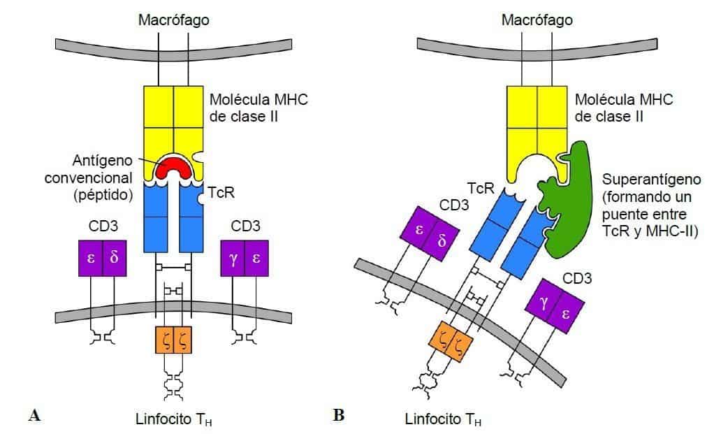
(A) Conventional antigens are presented to T cells on MHC class
II molecules. Recognition between MHC, the peptide and the TCR activates the number of TH cells specific for the antigen that is needed. (B) Superantigens are able to form bridges between the constant regions of MHC class II molecules and the TCR. There is no specific recognition between the superantigen, MHC and TCR, which activates an excessive number of TH cells and leads to a response that is dangerous for the individual. (Redrawn and modified from Regueiro JR et al. (2003) Inmunología. Biología y Patología del Sistema Inmune. (3ª Ed.) Editorial Médica Panamericana, Madrid.)
Conventional antigens presented on MHC class II molecules bind only to the TH cells that recognise them specifically, which represent a percentage far below 0.01% of the individual’s total T-cell repertoire. By contrast, superantigens are able to bind non-specifically to a multitude of MHC class II molecules and TCRs, which activates an excessive number of TH cells that, in this case, represents between 2% and 25% of the individual’s total T-cell repertoire.
Activation of TH cells leads to the production of cytokines, of which interleukin 2 (IL-2) is particularly noteworthy. With conventional antigens, cytokine production is a local event that allows the antigen to be eliminated appropriately. With superantigens, the overproduction of cytokines by the excessive number of activated TH cells cannot be contained locally and reaches the bloodstream (Figure 10.9). The entry of cytokines into the circulation produces adverse effects in the individual such as fever, nausea, vomiting and diarrhoea. In the most severe cases, shock, multiple organ failure, circulatory failure and even death may occur.
The ability of T-cell SAgs to bind non-specifically to large numbers of MHC class II molecules allows them to trigger life-threatening processes, which makes them a thoroughly undesirable group of molecules.
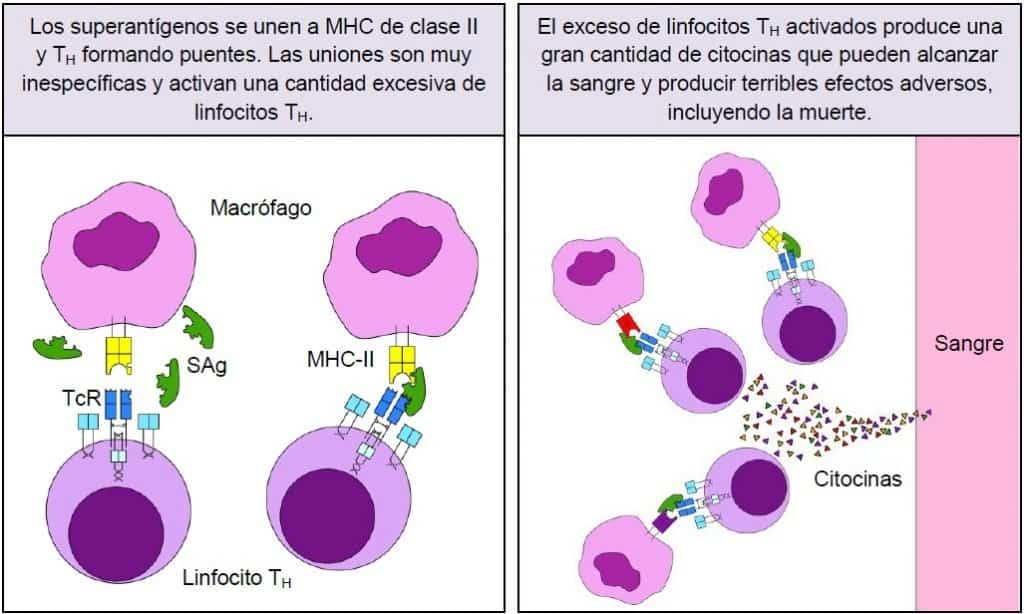
Superantigens bind to MHC class II and to the TCR, forming a bridge between the two structures. The non-specificity of the binding allows superantigens to activate up to 25% of the individual’s TH cells. The large quantity of cytokines produced by the activated TH cells cannot be contained locally and enters the bloodstream, producing undesirable effects.
(Courtesy of David Pérez Torres, University of Valladolid.)
A new class of superantigens has recently been described that specifically activates B cells and induces their apoptosis, and which are therefore known as B-cell superantigens. B-cell superantigens bind to the FAB regions of immunoglobulins outside their complementarity-determining regions. Staphylococcus protein A (SpA), the best-studied SAg of this class, reacts with the FAB regions of most VH3+ immunoglobulins, which are expressed in 30–60% of peripheral human B cells. Other B-cell SAgs are gp120 of human immunodeficiency virus (HIV), the Fv protein (a human hepatic sialoprotein) and Staphylococcus enterotoxin D (SED).
The ability of B-cell SAgs to bind to large quantities of serum immunoglobulin gives them potentially dangerous biological properties.
Presentation of non-protein antigens: the CD1 system
Numerous mechanisms give the immune system the ability to generate a great variety of T cells, which makes it possible to recognise a wide variety of antigens and mount an appropriate adaptive response against pathogens with very different characteristics. Most of the T-cell repertoire recognises antigenic peptides presented by the highly polymorphic MHC molecules. However, there is a significant population of T cells that do not react to antigens presented by MHC. Instead, this group of T cells binds to the molecules of an antigen-presenting family called CD1.
CD1 molecules are MHC class I-like glycoproteins (MHC class I-like) that are specialised in binding lipids and glycolipids for subsequent presentation to T cells. There are five CD1 genes on chromosome 1, called CD1A, CD1B, CD1C, CD1D and CD1E. Despite the limited allelic polymorphism of the CD1 system, these molecules are able to bind and present a wide range of lipid structures from pathogens and even from transplanted foreign tissues.
CD1 (Figure 10.10) is the first glycoprotein expressed by thymocytes during thymic differentiation. Structurally it is a heterodimer made up of an α chain, which contains three domains (α1, α2 and α3), with which a soluble protein called β2-microglobulin associates non-covalently. Between the α1 and α2 domains a physical groove forms that allows lipids to bind, called the antigen-binding groove (by analogy with the peptide-binding groove of the MHC system).
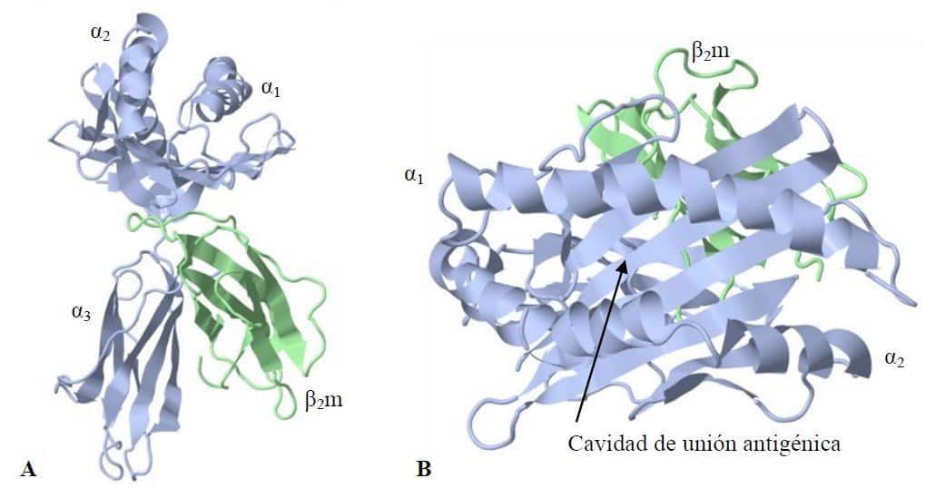
CD1 represents a third lineage of antigen-presenting molecules but, unlike MHC molecules, the type of antigen presented is lipid in nature. The crystal structure of mouse CD1d1, which corresponds to human CD1d, adopts a conformation very similar to that of MHC class I. (A) Note the structural similarities between MHC class I and CD1. CD1 is made up of an α chain with three domains and an associated β2-microglobulin molecule. As in MHC class I molecules, the α1 and α2 domains form the binding site for antigens. (B) The antigen-binding groove of CD1 is shown; it is significantly narrower
than the peptide-binding groove of MHC molecules but much deeper, and has two hydrophobic pockets. (RCSB PDB ID 1CD1)
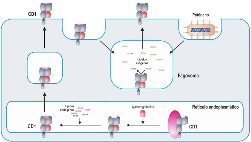
CD1 molecules are synthesised in the rough endoplasmic reticulum and then undergo a maturation process similar to that of MHC class I molecules (Figure 10.11). The α chains are glycosylated by glycosidases in the endoplasmic reticulum and their folding is mediated by chaperones such as calnexin, calreticulin and ERp57. The subsequent association of β2-microglobulin stabilises the complex and, in most molecules of the family, is an essential requirement for CD1 to be expressed on the cell surface.
(A) Conventional antigens are presented to T cells on MHC class
II molecules. Recognition between MHC, the peptide and the TCR activates the number of TH cells specific for the antigen that is needed. (B) Superantigens are able to form bridges between the constant regions of MHC class II molecules and the TCR. There is no specific recognition between the superantigen, MHC and TCR, which activates an excessive number of TH cells and leads to a response that is dangerous for the individual. (Reproduced from Regueiro J.R.,
López C., González S. & Martínez E. (2011) Inmunología. Biología y Patología del Sistema Inmune. (4ª Ed.) Editorial Médica Panamericana, Madrid.)
CD1 molecules are expressed only on professional antigen-presenting cells, especially dendritic cells and macrophages. CD1 molecules present glycolipid antigens to T cells, but not to conventional ones: to double-negative cells (CD4– CD8–). CD1d, which is also expressed on intestinal epithelium, presents its antigens to a special type of lymphocyte called NKT cells, which carry the characteristic markers of NK cells but are a specialised type of T cell, with cytotoxic function and the ability to synthesise cytokines.
The CD1 system is thought to be complementary to the HLA system; whereas HLA molecules present a sample of the peptides from inside the cell, CD1 molecules present lipids. In this way a healthy cell will present only self lipids, but an infected cell may present microbial lipids, which can then be recognised by T
cells. The lipid diversity of living organisms is much smaller than that of proteins or peptides; for this reason CD1 molecules are not polymorphic, as they are designed to accommodate a limited and well-defined number of lipids.