Here is the outline explained in the previous video. If you have any questions, post them in the comments section at the bottom of the page.
The MHC class II pathway
MHC class II molecules are heterodimers made up of an α chain and a β chain joined non-covalently. Both chains are synthesised independently in the endoplasmic reticulum. Alongside them, an invariant chain (I) protein, also called CD74, is synthesised; it stabilises the heterodimer and blocks the peptide-binding groove.
In this presentation pathway, the pathogen is taken up and internalised into an endosome (phagosome) by phagocytosis by a professional antigen-presenting cell. The phagosome containing the pathogen fuses with a primary lysosome loaded with hydrolytic enzymes and proteases (cathepsins). Their fusion gives rise to a secondary lysosome or phagolysosome in which, after the pathogen has been degraded, antigenic peptides of various lengths and sequences appear. Phagolysosomes empty their contents into an endosome unique to professional antigen-presenting cells, called the peptide-loading compartment (Figure 10.3, red arrows).
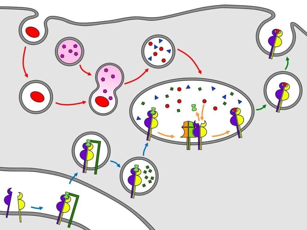
Extracellular antigens are degraded in a phagolysosome and sent to the peptide-loading compartment. MHC class II molecules synthesised in the endoplasmic reticulum are very unstable and therefore associate with CD74. MHC class II bound to CD74 travels to the peptide-loading compartment and, on the way, CD74 is hydrolysed except for a portion called CLIP, which blocks the peptide-binding groove. In the peptide-loading compartment, HLA-DM allows CLIP to leave and one of the antigenic peptides to enter the MHC class II molecule. The MHC class II molecule bound to its peptide is sent to the Golgi apparatus, where it undergoes glycosylation. Finally, the MHC class II molecule bound to its peptide is sent to the surface of the professional antigen-presenting cell. (Courtesy of David Pérez
Torres, University of Valladolid.)
MHC class II molecules blocked by CD74 migrate from the endoplasmic reticulum to the peptide-loading compartment, an organelle unique to professional antigen-presenting cells. During migration, CD74 is hydrolysed except for the portion that blocks the peptide-binding groove, called CLIP (Figure 10.3, blue arrows).
The peptide-loading compartment contains antigenic peptides from the phagolysosomes and MHC class II molecules blocked by CLIP. For antigen–MHC class II complexes to form, HLA-DM is required; this molecule acts as a peptide exchanger: it displaces CLIP and exposes the peptide-binding groove of the MHC class II molecule, allowing the antigenic peptides to bind. HLA-DM molecules differ from MHC class II molecules in that they are not polymorphic, do not associate with the invariant chain (I) (CD74), and are not expressed on the cell surface (Figure 10.3, orange arrows).
When one of the antigenic peptides binds to the peptide-binding groove for which it is specific, MHC class II completes its folding and migrates to the Golgi apparatus, where it undergoes glycosylation. After the necessary modifications, it is sent to the membrane of the professional antigen-presenting cell in an exocytic vesicle (Figure 10.3, green arrows). The peptide lodged in the peptide-binding groove of the MHC protein is presented to a T-cell receptor that recognises the peptide and the MHC chains simultaneously. If both structures are not recognised at the same time, the T cell is not activated.