Here is the outline explained in the previous video. If you have any questions, post them in the comments section at the bottom of the page.
Antigen presentation to T cells: overview
For antigens to be recognised by the TCRs of T cells, two consecutive phases are required:
- Antigen processing: the degradation of the protein antigen into peptides by intracellular digestion mechanisms.
- Presentation of the processed antigen: the association of some of the peptides obtained in the previous step with the peptide-binding groove of the molecules encoded by genes of the major histocompatibility complex (MHC).
Pathogens may originate inside or outside the cell (Figure 10.1). In the first case, the antigen is presented through the cytosolic pathway, which involves TC cells and MHC class I molecules; in the second case, it is presented through the endocytic pathway, which requires phagocytosis of the antigen and involves TH cells and MHC class II molecules (Table 10.1).
Most antigens can be presented to T cells by one of these two pathways, although some, such as lipid antigens, escape both.
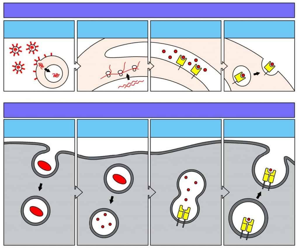
Depending on their origin, antigens can be presented by two pathways. Intracellular antigens, such as the genetic material of a virus, are presented on the surface by MHC class I molecules through the cytosolic pathway. Extracellular antigens, such as a bacterium or its remains, are presented on the surface by MHC class II molecules through the endocytic pathway. (Redrawn from Murphy K et al. (2008) Janeway’s Immunobiology (7th Ed.) Garland
Science, New York.)
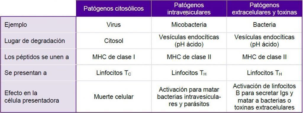
The table summarises the characteristics of the processing and presentation of different
pathogens. (Modified from Murphy K et al. (2008) Janeway’s Immunobiology (7th Ed.) Garland Science, New York.)
The MHC class I pathway
MHC class I molecules consist of an α chain associated with a soluble protein called β2-microglobulin; both are synthesised independently in the endoplasmic reticulum. The MHC class I protein is very unstable while it folds in the endoplasmic reticulum, so it associates with chaperone proteins.
At the start of folding, the α chain associates with a chaperone called calnexin. The subsequent binding of β2-microglobulin to the α chain requires calnexin to be replaced by calreticulin. This complex associates with tapasin and moves close to a heterodimer made up of TAP-1 and TAP-2. The function of the TAP complex is to transport peptides from the cytosol into the lumen of the endoplasmic reticulum, using ATP.
Viral proteins are degraded in the cytosol into peptides of various lengths and sequences by the proteasome, a multi-enzyme complex made up of four stacked rings, inside which lie subunits with proteolytic activity called LMP. These subunits, together with TAP-1 and TAP-2, are encoded by genes located in the MHC class II region.
Once the peptides are in the lumen of the reticulum, they fit at random into the peptide-binding groove of MHC class I molecules until they find the right binding site. When this happens, the MHC class I molecule completes its folding and is transported to the Golgi apparatus, where it undergoes glycosylation.
After undergoing the relevant modifications in the Golgi apparatus, the peptide–MHC complex is released to the cell surface in an exocytic vacuole, where it is anchored, waiting to be recognised by the TCR of a TC cell (Figure 10.2).
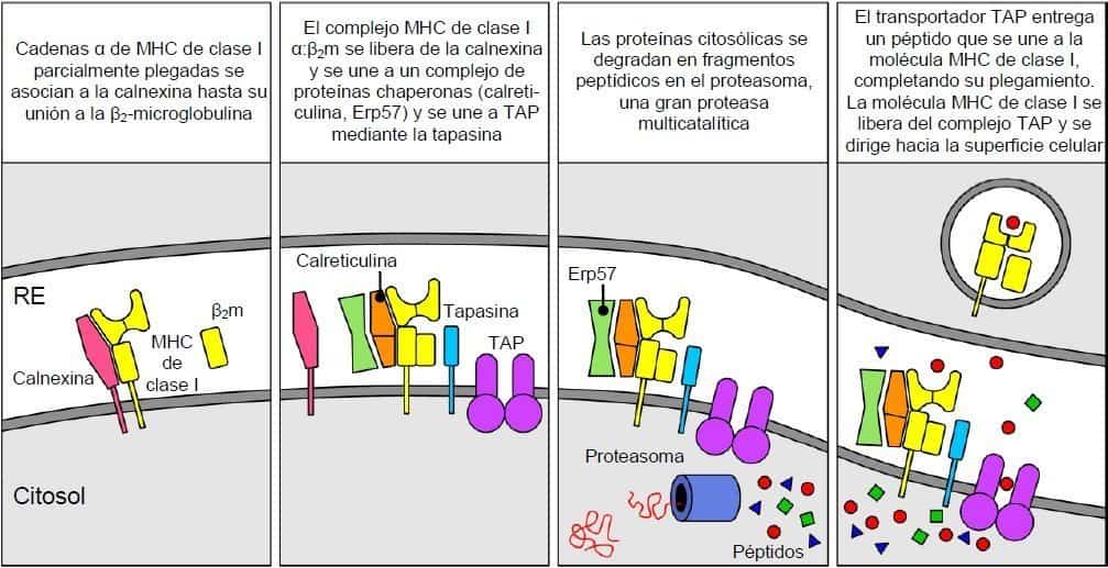
Note that MHC class I molecules do not leave the endoplasmic reticulum (ER) until they have bound a peptide. Once the peptide has bound to the MHC molecule, the peptide–MHC complex leaves the reticulum and is sent to the Golgi apparatus, where it may undergo modifications. The complex is then sent to the cell surface. (Redrawn and modified from Murphy K et al. (2008) Janeway’s Immunobiology (7th Ed.) Garland Science, New York.)