Although clinical detection of these conditions takes place in the vast majority of cases in childhood, it can also occur in adults. There is a “list of ten warning signs for detecting these conditions” that was created by the Jeffrey Modell Foundation. It is a list of ten clinical signs that should make us think of an immunodeficiency.
Items on the list:
- Eight or more ear infections (“otitis”) in one year or less.
- Two or more severe “sinus infections” in one year or less.
- Two months (or more) of antibiotic therapy with little or no effect.
- Two or more pneumonias in one year.
- Difficulty gaining weight or delayed growth (problems gaining or losing weight or height).
- Recurrent abscesses: deep skin abscesses (or abscesses of other organs).
- Persistent thrush in the mouth (or on the skin) in children over 1 year of age.
- Need for intravenous antibiotics to clear infections.
- Two or more deep-seated infections: cellulitis, meningitis, osteomyelitis or sepsis.
- Family history of primary immunodeficiency.
The presence of one of these symptoms should already set off alarm bells, and it is advisable to investigate the possible presence of primary autoimmune disease. If more than one of these symptoms is present, the diagnosis of immunodeficiency is already clear.
Typical general clinical features of immunodeficiencies
- Increased risk of infections.
- Increased risk of tumours: the main tumours are non-Hodgkin lymphoma and gastric cancer, because in some immunodeficiencies lymphocytes do not die or there is less control.
- Increased risk of autoimmune diseases (especially in IgA deficiency).
- Increased risk of atopic diseases (especially in IgA deficiency).
- Frequent abnormalities of growth (weight and height).
A patient arrives with a clinical picture that makes us suspect an immunodeficiency. We look at the family history. If there are no cases, it may be a secondary immunodeficiency.
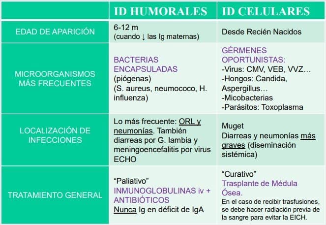
A mixed picture of immunodeficiency with other diseases can appear. The bacteria or viruses that appear in a child with an immunodeficiency (ID) can give us a clue to the type of ID the child has:
Diagnosis of PIDs
- Clinical diagnosis: family history of immunodeficiencies, no response to antibiotics, recurrent infections…
- A basic screening is carried out: tests of the kind we do in the practical classes, such as quantification of the predominant lymphocyte subpopulations and quantification of immunoglobulins (IgG, IgM and IgA). A basic blood count can also be done, in which we look at the number in each lymphocyte group and do a leucocyte count.
- If the parameters obtained in the test are normal, only a few options remain, related to complement and innate immunity. We will move on to more specific tests, so the patient will have to be sent to a reference centre.
- If they are abnormal, there is a clear immunodeficiency, and all that remains is to determine whether it is humoral or combined cellular. This is not something that just any doctor does either; the patient is sent to a reference centre.
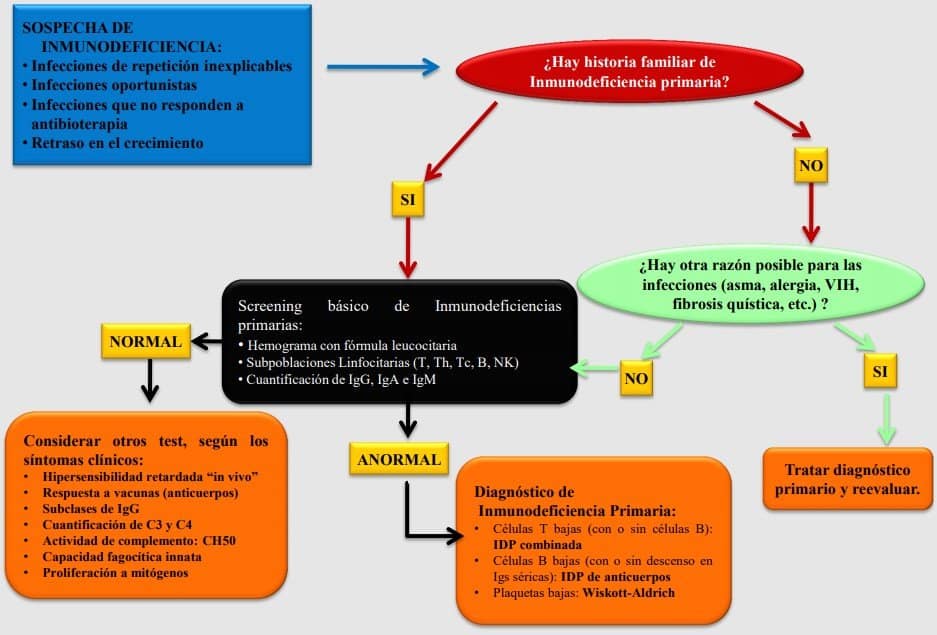
A patient comes to the clinic with a suspected immunodeficiency. First we see whether there is a family history of primary immunodeficiency. This is a key piece of information: if there is none, the possibility of a primary immunodeficiency is ruled out, so it would be secondary. We look for possible causes of the symptoms our patient presents and, if we find nothing, we do a basic screening. This is done using various methods (lymphocyte subpopulations (T, B, NK), immunoglobulin quantification, blood counts…). If everything is normal, we still cannot rule out an immunodeficiency, and the sample is taken to a specialised laboratory that is specific for those parameters, so as to find out exactly what the patient has.
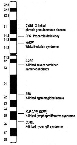
Example of a PID: X-linked agammaglobulinaemia (Bruton syndrome).
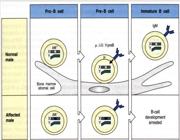
The defective gene is the Btk gene (B-cell tyrosine kinase). Btk is needed to rearrange the light chain in B cells at the pre-B stage and express it on the outside of the membrane together with the heavy chain. (B lymphocytes mature in the bone marrow and, to pass from the pre-B stage to the immature B-cell stage, they have to rearrange the light chain, for which Btk is necessary.)
Patients with Bruton syndrome (oBTK) cannot complete B-cell maturation, and their cells remain at the pre-B stage. The pre-B cell does not progress to an immature B cell, so it keeps a surrogate light chain (λ5). They usually have no B cells in the blood, and the few they do have are at the pre-B stage, with a heavy chain but no light chain, so they do NOT produce immunoglobulins.
It appears only in males: it is sex-linked.
Clinical features:
- Early onset (in the first year) of respiratory and/or intestinal infections: from the moment maternal IgG disappears. This is from weaning onwards or, in the case of formula feeding, from the very first moment of life.
- Severe bacterial and viral infections of deep organs.
- Oedema and arthritis (septic or not) in all joints.
- Neutropenia, alopecia and malabsorption (not in all cases).
These children have no immunoglobulins or B lymphocytes. It is a disease that presents very early with infections of the respiratory and gastrointestinal tract. This usually happens when breastfeeding ends or, if there is none, earlier. The infections are caused by both bacteria and viruses and are usually accompanied by arthritis and joint inflammation (joint oedema); the children also show growth retardation. There are other symptoms that do not appear in all cases, such as neutropenia.
Laboratory findings:
- Serum levels of IgG, IgA and IgM very low or undetectable (0).
- Natural antibodies: absent.
- Vaccine antibodies: absent.
- Circulating B cells: <2% (pathognomonic sign, specific to the disease (BTK)).
The screening test, or definitive test, is serum immunoglobulin quantification, which will show low or totally absent levels. If this is the case, lymphocyte detection by flow cytometry must be done and the lymphocyte populations checked.