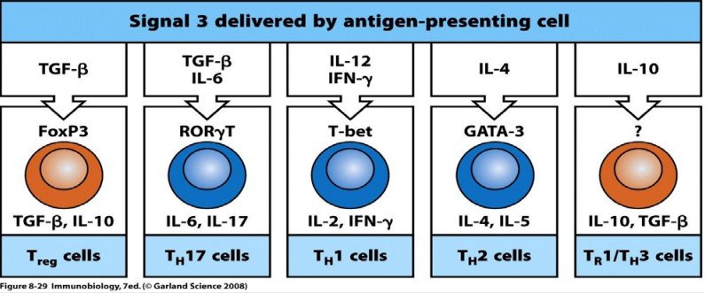
Differentiation of CD4+ T cells
T-cell lymphopoiesis takes place in the thymus. Both CD4 and CD8 cells are produced, but here we will focus on the CD4 cells.
APCs and the cytokines they produce, together with the pathogen and the location (environment), are decisive for the differentiation of T cells.
The immune system is able to adapt and select the best (most effective) mechanism, depending on the type of pathogen it has to face.
Within the thymus itself:
- Some of the CD4 cells will already differentiate into regulatory T cells. These are called natural or central regulatory T cells (nTreg).
- The rest, the vast majority, leave into the circulation as naive effector T cells (Th0)
The next differentiation will take place in the peripheral lymphoid organs. Whether the cell differentiates into one type or another will depend on the microenvironment and on the dendritic cell presenting the antigen.
In general, after activation they differentiate into the effector T cells Th1, Th2 and Th17. But a few, under certain conditions provided by the dendritic cell and the microenvironment, can differentiate into regulatory cells, which are called iTreg (inducible or induced regulatory cells).
Depending on the antigen, the response will be different:
1- When an intracellular pathogen enters:
- The antibody-mediated defence mechanism is NOT effective. Macrophages are the first to take up the microorganism. When the macrophage is activated and phagocytoses the pathogen, it secretes a series of substances, among them IL-12, which promotes the differentiation of the Th1 cells that have recognised the antigen.
- Th1 cells activate the cells responsible for innate and adaptive immunity: they secrete IFN-γ, which activates macrophages, NK cells and CD8+ cytotoxic T cells.
2- When the pathogen is extracellular (the microorganism is in the plasma or serum)
- In this case the lymphocytes secrete IL-4 (in the absence of IL-12). IL-4 induces the differentiation of Th2 cells. These cells produce IL-5, IL-10 and IL-13, which induce the production of IgG and IgM antibodies in B cells.
- They are also responsible for the activation of eosinophils: these are very effective cells for destroying extracellular organisms such as parasites and protozoa.
These two subpopulations are very effective and carry out a characteristic response. Their differentiation implies the inactivation of the other subtype, so that only the response appropriate to the situation is maintained.

The microenvironment
The microenvironment depends on the cells of the innate immune response that were activated first. It will be different if they are mast cells than if they are NK cells. In turn, the type of cells that were activated earlier depends on the type of infection.
- Intracellular infections, whether viral or bacterial, will normally provoke Th1 responses.
- Extracellular infections by bacteria and fungi will induce Th17 responses.
- Extracellular infections by large parasites (worms) will induce Th2 responses.
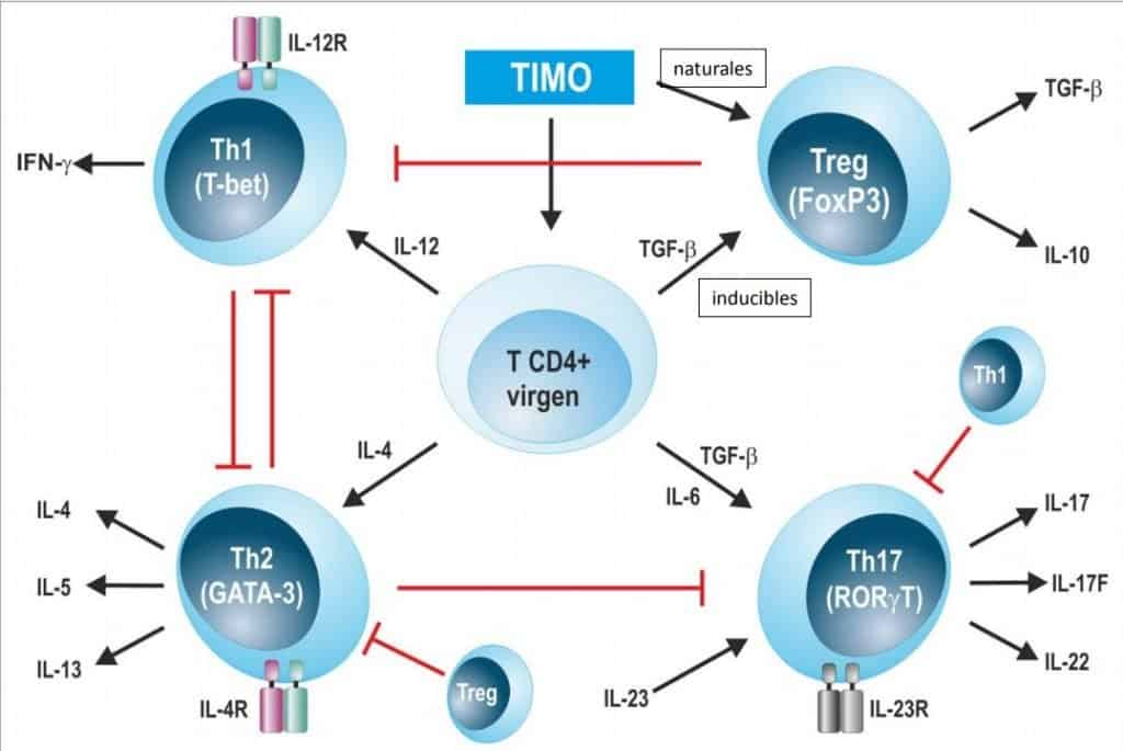
In the absence of antigenic stimulation in the lymph nodes, the cytokine that prevails in all environments is TGF-β. When TGF-β is the predominant cytokine, antigen presentation will always determine the differentiation of regulatory T cells. These produce two cytokines, the only ones that have a net inhibitory effect: IL-10 and TGF-β. These cells therefore circulate through the body and are responsible for keeping the immune system at rest, which is the best way to face the arrival of a pathogen. They differentiate through the FoxP3 signalling pathway. There are two types of Treg:
- Some leave the thymus already differentiated in regulatory form (natural signals): these are the CD4+CD25+ cells.
- Most differentiate in the periphery (inducible signals): these are the Tr1 and Th3 cells.
Th1 cells
- They are characterised by expression of the IL-12 receptor on their membrane (they differentiate in the presence of IL-12 and the absence of TGF-β). The CD4+ T cell differentiates into a Th1 cell in the presence of IL-12 (which induces expression of the IL-12 receptor); IL-12 binds to the receptor and a signalling pathway called T-bet is activated (each cytokine activates a different intracellular transduction pathway). The Th1 cell produces IFN-γ.
Th2 cells
- They are characterised by membrane expression of IL-4R (they differentiate in the presence of IL-4 and the absence of TGF-β). The binding of IL-4 to its receptor induces the signalling pathway called GATA-3. The Th2 cell releases IL-4, IL-5 and IL-13.
The Th17 cell
- It is activated in the absence of IL-12 and IL-4 and in the presence of IL-6 and TGF-β. In addition, IL-23 is present and interacts with its receptor. The transcription signals that are activated are those of RORγT.
When a great deal of IFN-γ is produced, the Th1 response is what is wanted rather than any other cell type, so IFN-γ is a blocker (inhibitor) of Th2 cells and of every other cell type. Thus, when Th1 cells are activated, by releasing IFN-γ they block the other cells, since at that moment the appropriate response is against intracellular pathogens. When the causative agent disappears, balance is restored and there are cells of every type.
(Each lymphocyte can be identified by its membrane receptors, by its intracellular signal transduction systems or by the expression of a series of cytokines.)
Subtypes of CD4+ T cells
- Th1 cells: they produce mainly two cytokines: IL-2, IFN-γ and TNF-β
- They activate macrophages and NK cells by releasing IFN-γ
- They help CD8+ T cells by releasing IL-2, which activates the cytotoxic cells, and these destroy the infected cell
- They help B cells in antibody production.
- Th2 cells: they synthesise IL-4, IL-5, IL-6 and IL-10.
- They help B cells in antibody production
- They activate mast cells and eosinophils
- Th17 cells: they secrete IL-17, IL-6, IL-23 and TGF-β
- They regulate the mobility of neutrophils and contribute to the integrity of the epithelium (they are responsible for acute inflammatory responses).
For many years Th1 cells had been linked to chronic inflammation, especially of the intestine (e.g. Crohn’s disease). However, after inhibiting the production of IL-12 some patients continued to have symptoms, and the presence of IL-23 was detected, so Th17 cells were also implicated.
- Treg cells: two types, which synthesise inhibitory cytokines:
- Those that leave the thymus directly as regulatory cells: nTregs (natural regulatory cells, CD4+ CD25+)
- Those that differentiate into regulatory cells in the periphery: Tr1 and Th3,
- Tfh: follicular helper T cells
- Th0: naive T cells