Immunocompetence is the ability to recognise an antigen specifically. This ability is acquired in the bone marrow.
Naive lymphocytes mature and differentiate in the bone marrow. From there they enter the circulation and reach the secondary lymphoid organs. The cells are constantly circulating, since they are naive cells and have a high capacity to move (activated cells create mechanisms to stay where they were activated). The antigens of the pathogens that have infected the body reach the secondary lymphoid organs.
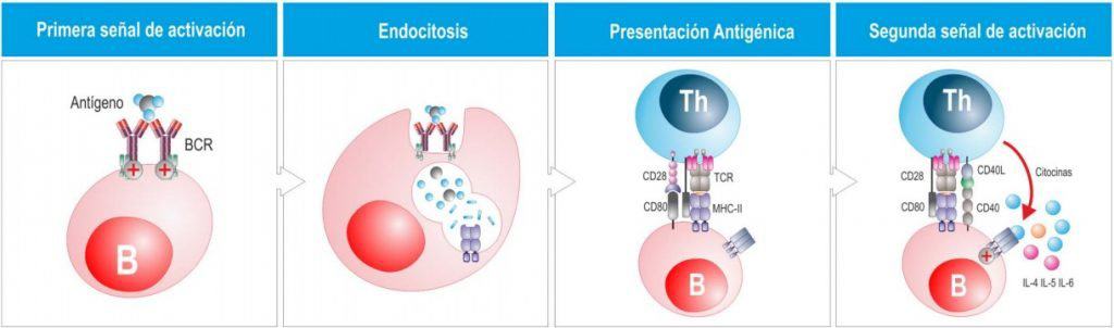
Bacteria, viruses and other pathogens, including tumour cells, express foreign antigens on their membrane. The B cell’s BCR receptors interact with their specific Ag.
After the B cell interacts with the antigen, it proliferates and creates a clone of lymphocytes with the same BCR. This first contact with the antigen (through an epitope) leads to partial activation of the B cell, which expresses, among other molecules, cytokine receptors and accessory molecules.
The B cell then behaves like a phagocyte and internalises the pathogen. It degrades and processes it and presents it to T cells on MHC molecules. The B cell will then act as an antigen-presenting cell. The difference from other antigen-presenting cells is that the specificity of B cells for antigens is much higher, so they interact with antigens at much lower concentrations. This, together with the interaction of CD28 with CD80/86 on the B cell, activates the T cell.
This interaction between T cells and B cells allows the second activation signal of the B cells to occur. The T cell expresses CD40L, which interacts with CD40 on the B cell, and this stimulates the pathway that forms plasma cells → it converts B cells into plasma cells so that they synthesise antibodies. The cytokines released by the T cell are responsible for the production of one Ig or another.
Th cells (CD4+) that recognise the peptide presented by the B cell’s MHC class II are activated and begin to proliferate and differentiate in order to activate the B cells. During antigen presentation a series of interactions take place between the B cells and the Th cells. Once differentiated, effector Th2 cells do not leave the secondary lymphoid organs, as Th1 or Tc cells do, but stay in the lymphoid organs and send signals that activate and expand the clone of B cells specific for the antigen that had been partially activated. The cytokines released by the Th2 cell indicate the isotype switch and also the type of pathogen present in the environment.
The B cell acts in two ways:
- Direct membrane-to-membrane contacts through a CD40 - CD40L molecule pair (part of the second stimulatory signal together with the cytokines).
- Via soluble factors (IL-4, IL-10, IL-13).
Depending on the type of cytokine, different isotypes will be produced. All this takes place in the lymph node.
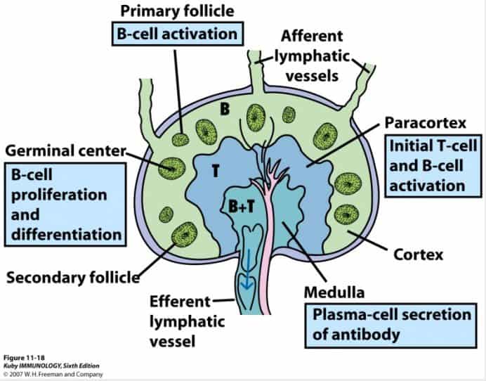
In the lymph nodes, activated B cells proliferate in the germinal centres, located in the cortex.
Primary lymphoid follicle
The first clone of activated B cells, which are proliferating. After a time (about a week), the primary follicle becomes a secondary lymphoid follicle, which has a denser central zone called the germinal centre, where we find dividing B cells, and a less dense peripheral zone made up of non-proliferating B cells, called the mantle. The B cells that proliferate in the germinal centre change their morphology and are distinguished from resting ones because they are larger. Centroblasts are the B cells that have recognised Ag and are dividing, and the B cells resulting from the previous division are the centrocytes, which have already stopped dividing. On the outside there is a layer of T cells.
The proliferating cells undergo a series of changes, improving their affinity (somatic hypermutation) and their effectiveness against the pathogen.
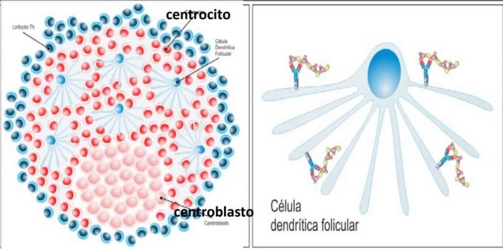
Follicular or interdigitating dendritic cells
Within the germinal centre. They present antigens (from microorganisms or bacteria) attached to their dendrites through opsonisation. What these cells pick up are immunoglobulin molecules that are in turn bound to an antigenic peptide. They have complement receptors and receptors for the Fc fragment of Ig, which allows these cells to present, in that germinal centre, all the foreign antigens that have reached the lymph node. They do not function as ordinary dendritic cells, since they are not antigen-presenting cells but act as an antigen display.
In the germinal centre a series of very important events take place that are needed to understand how the humoral immune response is formed.
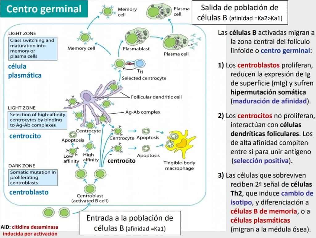
The B cells that have been activated migrate to the central zone of the lymphoid follicle, or germinal centre (they express BCR receptors and Ig on the membrane). They are organised around a germinal centre in different stages of differentiation.
- B cells that have just interacted with an antigen have affinity to bind to it, but it is not sufficient (low affinity): these are centroblasts (dividing cells located in the “dark zone”). Centroblasts proliferate and reduce the expression of Ig on their membrane.
- Centroblasts undergo a process known as somatic hypermutation (affinity maturation) (only in the lymph nodes!): the enzymes activation-induced cytidine deaminases induce a series of mutations that affect the hypervariable portions of the immunoglobulin molecule. They convert a cytosine into uracil and the variable region is restructured. The mutations may or may not be productive, but if they are productive they will improve affinity.
- Only those centroblasts that have undergone productive somatic hypermutation will survive. These centroblasts differentiate into centrocytes, which have an Ig with greater affinity for the specific antigen. The centrocytes move into the light zone, a zone where high-affinity selection takes place. Centrocytes do not proliferate but interact with the antigen-antibody complexes of the follicular dendritic cells. This is the process of positive selection, in which only those cells with maximum affinity survive.
- The cells that survive positive selection receive the second signal from TFH cells for isotype switching and differentiation into memory B cells or plasma cells (which migrate to the bone marrow). This signal comes from cytokines released by the T cell, which determines the type of Ig to be synthesised.
- Plasma cell producing immunoglobulins: it does not express immunoglobulin molecules on its membrane. It is already activated and needs neither to divide nor to undergo somatic hypermutation. Plasma cells head for the bone marrow. They are cells with a large RER that produce antibodies. They are activated according to which cells are needed.
- Memory cells: they do express immunoglobulin molecules on their membrane, that is, they do recognise Ag. They will not divide. These cells will recirculate again and in many cases will become lodged in tissues. They are responsible for secondary responses.
In the end we have two types of cells with much more affinity than they had at the start.