Here is the outline covered in the previous video. If you have any questions, post them in the comments section at the bottom of the page.
Regulation of the complement system
Given the devastating destructive effects triggered by the complement system, and the way its activation is rapidly set off and amplified, it is not surprising that there are multiple mechanisms to prevent uncontrolled activation.
A passive mechanism for regulating the complement system is the lability of some of its components, which allows them to be inactivated spontaneously when they are not stabilised in a reaction with other components. For example, the C3 convertase of the alternative pathway has an active half-life of about 5 minutes unless it is stabilised by properdin.
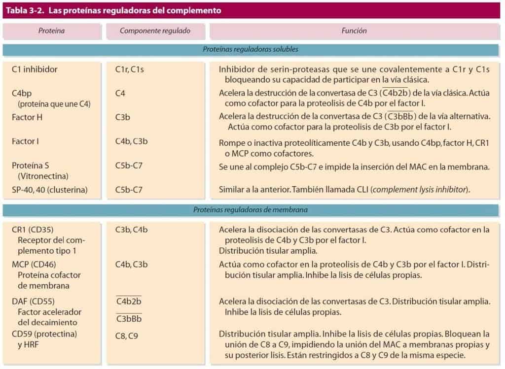
Active mechanisms for regulating the complement system include a set of regulatory proteins that inactivate different complement elements. Complement regulatory proteins can be found in soluble form or anchored to the membrane, as detailed in Figure 12.8.
It is necessary to distinguish between complement receptors and the complement regulatory proteins that coat cell membranes. In both cases their ligands are complement components, but their biological functions differ: the former help cells of the immune system to improve the elimination of pathogens, while the latter inhibit uncontrolled complement activation on healthy tissues. There is one exception: CR1 does both.
(Reproduced from Regueiro J.R., López C., González S. & Martínez E. (2011) Inmunología. Biología y Patología del Sistema Inmune. (4th Ed.) Editorial Médica Panamericana, Madrid.)
Regulation of complement before convertase assembly
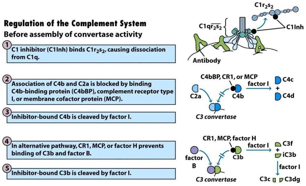
C1 inhibitor (C1Inh) is a soluble glycoprotein that can form a complex with C1r2s2, causing dissociation of C1q and thereby preventing activation of C4 or C2 (Figure 12.9-1).
The reaction catalysed by C3 convertase from any of the activation pathways can generate hundreds of molecules of C3b. These, in turn, can bind to nearby cells and damage them by inducing MAC formation on them. As a regulatory mechanism, C3b undergoes spontaneous hydrolysis once it moves more than 40 nm away from the C3 convertases. In addition, there are a number of proteins called RCA (regulators of complement activation) that regulate the activity of the C3 convertases. They include C4b-binding protein (C4bBP), complement receptor 1 (CR1) and membrane cofactor protein (MCP). Each of these regulatory proteins binds to C4b and prevents it from binding to C2b. After this, another regulatory protein, factor I, cleaves C4b into bound C4d and soluble C4c (Figure 12.9-2,3).
A similar regulatory sequence prevents assembly of the C3 convertase of the alternative pathway. In this case, CR1, MCP or a regulatory component called factor H bind to C3b and prevent it from binding to factor B. Factor I then cleaves C3b into a bound iC3b fragment and a soluble C3f fragment. Further proteolysis of iC3b by factor I gives rise to soluble C3c and membrane-bound C3dg (Figure 12.9-4,5).
(Reproduced from Kindt T.J., Goldsby R.A., Osborne B.A. & Palacios R. (2007) Inmunología de Kuby (6th Ed.) McGraw-Hill Interamericana, Madrid.)
Regulation of complement after convertase assembly
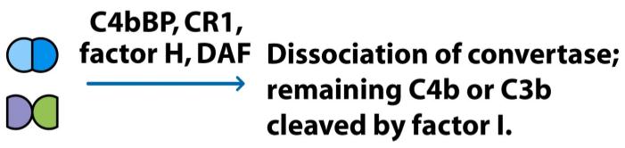
The RCA proteins, particularly C4bBP, CR1 and factor H, also act on the assembled C3 convertase and cause it to dissociate. Decay-accelerating factor (DAF) or CD55, a membrane glycoprotein, can also dissociate the C3 convertase. After dissociation of the C3 convertase, factor I cleaves the remaining membrane-bound C4b or C3b component, irreversibly inactivating the convertase (Figure 12.10).
(Reproduced from Kindt T.J., Goldsby R.A., Osborne B.A. & Palacios R. (2007) Inmunología de Kuby (6th Ed.) McGraw-Hill Interamericana, Madrid.)
Regulation of complement at MAC assembly
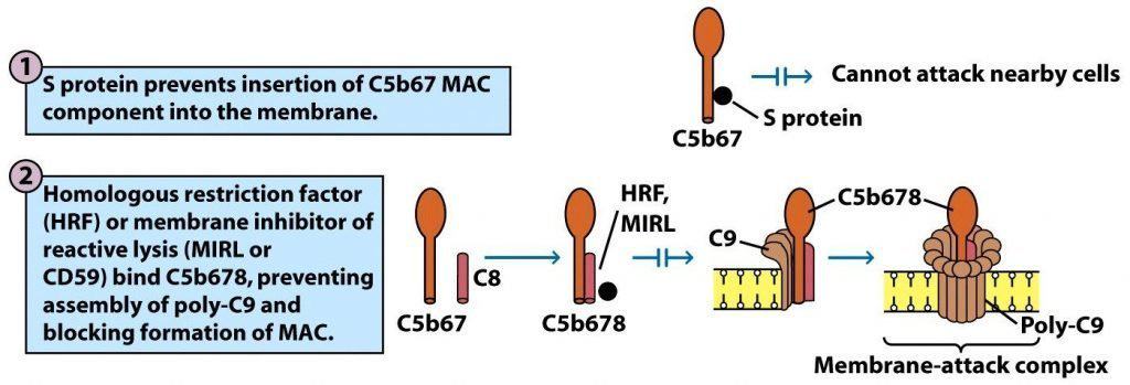
Formation of the C5b67 complex carries a high risk of lysis for healthy host cells. For this reason, there are multiple serum proteins that counter this threat by binding to the complex and preventing its insertion into the membrane of nearby cells. Thus, protein S can bind to C5b67 and prevent its insertion into the cell membrane (Figure 12.11-1).
Complement-mediated cell lysis is more efficient when the complement comes from a species different from that of the cells being lysed. This phenomenon is attributed to the so-called homologous restriction factor (HRF), membrane inhibitor of reactive lysis (MIRL), CD59 or protectin. This protein is found in the membrane of many cell types and protects cells from non-specific complement-mediated lysis by binding to C8, thereby preventing assembly of poly-C9 and its insertion into the membrane. This inhibition occurs only when the complement components come from the same species as the target cells (Figure 12.11-2).
(Reproduced from Kindt T.J., Goldsby R.A., Osborne B.A. & Palacios R. (2007) Inmunología de Kuby (6th Ed.) McGraw-Hill Interamericana, Madrid.)