Here is the outline covered in the previous video. If you have any questions, post them in the comments section at the bottom of the page.
Effector mechanisms of the complement system
Once the complement system has been activated by any of the possible pathways, its components interact in a tightly regulated cascade to carry out four basic functions (Figure 12.1):
- Lysis of Gram-negative bacteria, parasites, enveloped viruses and host cells.
- Opsonisation of particulate antigens, which allows them to be phagocytosed.
- Activation of the inflammatory response, through complement receptors and the secretion of immunoregulatory molecules.
- Clearance of immune complexes (immune clearance), which removes antigen-antibody complexes from the circulation via the spleen and liver.
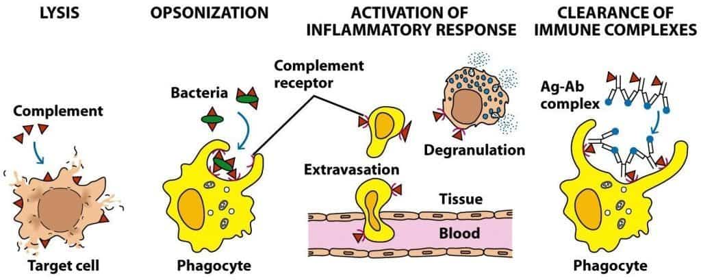
(Reproduced from Kindt T.J., Goldsby R.A., Osborne B.A. & Palacios R. (2007) Inmunología de Kuby (6th Ed.) McGraw-Hill Interamericana, Madrid.)
Lysis of Gram-negative bacteria, parasites, enveloped viruses, erythrocytes and nucleated cells
Formation of the membrane attack complex (MAC) damages the wall of Gram-negative bacteria, parasites, enveloped viruses and host cells (erythrocytes and nucleated cells) by forming a pore in their lipid bilayer. Although this effector mechanism of complement may seem highly destructive, its role in host defence is in fact limited to a few pathogens, and the mechanisms of opsonisation and activation of the inflammatory response are far more important.
MAC formation is the main defensive weapon of complement against Gram-negative bacteria, especially those of the genus Neisseria. These bacteria are associated with gonorrhoea, a sexually transmitted disease, and with some forms of bacterial meningitis, a serious infection of the central nervous system. MAC formation damages the structural elements of the bacterium, causing an osmotic shock that leads to its lysis. However, some groups of Gram-negative bacteria have developed mechanisms to escape the action of the MAC. For example, some strains of Escherichia coli or Salmonella have developed side chains on the lipopolysaccharide of the cell wall that prevent the MAC from inserting into the bacterial wall. Patients with deficiencies of complement components C5-C9 are more susceptible to the diseases mentioned above. In general, Gram-positive bacteria are resistant to the action of the MAC, thanks to their thick peptidoglycan layer and the bacterial capsule.
The outermost layer of virions, or viral particles, may be a capsid or an envelope. Enveloped viruses are more labile in the face of environmental factors such as acid, detergents, desiccation or heat, whereas non-enveloped viruses, contrary to what might be expected, are resistant to all of these factors. Most enveloped viruses are susceptible to complement-mediated lysis. The viral envelope is derived mostly from the plasma membrane of infected host cells, so MAC-mediated pore formation is possible. Complement and antibodies play a key role in defence against viruses and are crucial for limiting viral spread during acute infection and for protecting against reinfection. Viruses susceptible to complement-mediated lysis include the families Herpesviridae (HSV-1, HSV-2, VZV, EBV, CMV, etc.), Orthomyxoviridae (influenza A, B and C viruses), Paramyxoviridae (measles virus, mumps virus, respiratory syncytial virus, etc.) and Retroviridae (HIV, HTLV, etc.).
Host cells can also be lysed by the MAC, as occurs with erythrocytes, which have a limited capacity to resist and repair damage to their membrane, in processes such as autoimmune haemolytic anaemia and other haemolytic disorders. Nucleated cells, on the other hand, can escape the lytic action of the MAC more easily. Even so, the formation of pores in the membrane allows Ca2+ ions to enter the cell, activating numerous mechanisms that can contribute to disease. The mechanism by which nucleated cells escape the action of complement is endocytosis of the MAC, so that the cell can repair the damage to its membrane and restore its osmotic balance. An unfortunate consequence of this is that complement-mediated lysis triggered by antibodies specific for tumour cell antigens, which is a possible cure for cancer, is ineffective because these cells endocytose the MAC.
Opsonisation
Activation of the complement system and its corresponding amplification mechanisms results in the coating of the surface of target elements, such as bacteria and immune complexes, by opsonins (the larger fragments) resulting from proteolysis of the inactive molecules.
The main factor responsible for complement-mediated opsonisation is C3b, although iC3b and C4b also have this function. Phagocytic cells express complement receptors (CR1, CR3 and CR4) able to bind opsonins. Binding between opsonins and their receptors allows phagocytosis of the opsonised element, which may be intact or in particulate form after lysis, starting antigen processing and accelerating the synthesis of specific antibodies (Figure 12.2).

(Reproduced from Kindt T.J., Goldsby R.A., Osborne B.A. & Palacios R. (2007) Inmunología de Kuby (6th Ed.) McGraw-Hill Interamericana, Madrid.)
Activation of the inflammatory response
During formation of the MAC, small peptides are generated by cleavage of the inactive complement proteins. These fragments, called anaphylatoxins (C3a, C4a and C5a), bind to receptors on mast cells and basophils, inducing their degranulation and the consequent release of histamine, TNF-α and other inflammatory mediators responsible for starting the inflammatory response (Figure 12.3).
In addition, anaphylatoxins induce smooth muscle contraction and increase vascular permeability, allowing plasma laden with antibodies and phagocytic cells to enter the site where the antigen is located.
C3a and C5a can induce adhesion of monocytes and neutrophils to the endothelium, allowing their extravasation and migration towards the site of complement activation in a tissue. The anaphylatoxin C5a is the most potent mediator of these processes and is effective at picomolar concentrations. C5a can also induce expression of the CR1 receptor on phagocytic cells, facilitating phagocytosis of antigens coated with C3b.
Anaphylatoxins are highly reactive substances and are regulated by a serum protease called carboxypeptidase N, which cleaves an arginine residue from the carboxyl-terminal end of the molecules and produces the corresponding desarginated forms. The des-Arg form of C3a is completely inactive, whereas the des-Arg form of C5a retains some chemotactic activity and the ability to contract smooth muscle.

(Reproduced from Male D., Brostoff J., Roth D.B. & Roit I. (2013) Immunology (8th Ed.) Elsevier Saunders, Philadelphia.)
When anaphylatoxins are produced in large amounts or injected systemically, they induce generalised circulatory collapse, which produces a syndrome resembling shock mediated by IgE-type antibodies. This reaction is called anaphylactic shock, and it is a very severe allergic reaction that can cause death by asphyxia within a few minutes (Figure 12.4). Treatment must be started immediately whenever it is suspected, by injecting 0.3-0.5 mg of intramuscular adrenaline (in adults), which can be repeated every 5-10 minutes, and an antihistamine such as dexchlorpheniramine (Polaramine®). Appropriate ventilatory and haemodynamic support measures must also be provided.
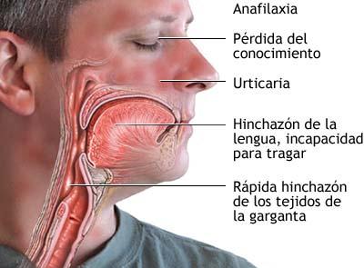
(Reproduced from ADAM.)
Clearance of immune complexes
Immune complexes are complexes formed by antibodies and remnants of pathogens or dead host cells. Because of their marked tendency to aggregate and incorporate further components, immune complexes can be deposited in capillary beds, the skin, the kidney and other organs, causing disease. This occurs in diseases such as systemic lupus erythematosus.
Activation of the classical complement pathway efficiently opsonises immune complexes and prevents them from precipitating in the tissues. To do this, C3b coats the antigens at the core of the immune complex, preventing its growth. In addition, C3b breaks up large immune complexes, preventing antigen-antibody interaction. Finally, the C3b and C4b coating the surface of the immune complexes interact with, and bind to, the CR1 receptors on erythrocytes. Adhesion to erythrocytes allows the dangerous immune complexes to be transported to the macrophages of the spleen and liver, where they are internalised and destroyed.
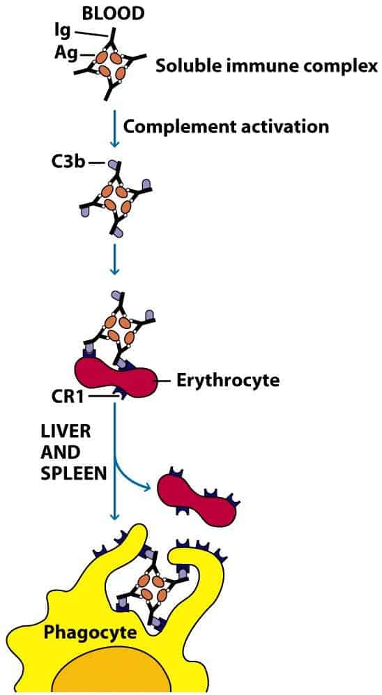
(Reproduced from Kindt T.J., Goldsby R.A., Osborne B.A. & Palacios R. (2007) Inmunología de Kuby (6th Ed.) McGraw-Hill Interamericana, Madrid.)