Here is the outline explained in the previous video. If you have any questions, post them in the comments section at the bottom of the page.
Basic concepts and nomenclature
When a pathogen crosses the epithelial barrier and breaches the individual’s first line of antimicrobial defence, the first component it encounters is an element of innate immunity known as the complement system, or simply complement.
Complement is a collection of soluble proteins present in the blood and body fluids. More than 30 proteins belong to complement and they are produced mainly in the liver. In the absence of infection, these proteins circulate in an inactive form. In the presence of pathogens, or of antibodies bound to pathogens, the proteins of the complement system become activated.
Complement proteins interact with one another in specific ways through different activation pathways, the ultimate consequence of which is the death of the pathogen. This is achieved by facilitating phagocytosis and by inducing an inflammatory response able to fight the infection. There are three pathways of complement activation: the classical pathway, which is triggered by antibodies attached to the surface of the pathogen, the alternative pathway, which is triggered by the mere presence of the antigen, and the lectin pathway, which is activated by molecules on the pathogen surface (Figure 11.1). All of them converge on the formation of the C3 convertase.
The proteins of the complement system are synthesised in the liver as zymogens, or proenzymes, that is, in an inactive form, and require proteolytic cleavage to become active. As a general rule, each protein of the complement system is activated by another protein of the system. The proteolytic cascades generated in this way are the origin of the activation pathways, and are responsible for generating the effector components of complement (the common terminal pathway) and for amplifying the response, which allow the pathogen to be eliminated.
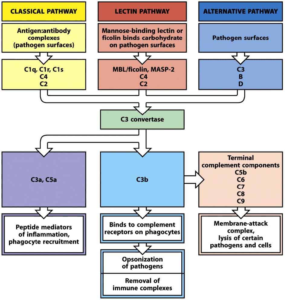
(Reproduced from Murphy K et al. (2012) Janeway’s Immunobiology (8th Ed.) Garland Science, New York.)
Nomenclature
The nomenclature of the proteins of the complement system can be confusing. The first complement activation pathway to be discovered was the classical pathway, whose proteins are designated by the letter C and a number (C1, C2, C3, C4, C5, C6, C7, C8 and C9). This number does not describe the order in which the components interact, but the order in which the molecules were discovered. When a protease cleaves a complement protein to activate it, two products result. The products of this reaction are designated with a suffix consisting of a lower-case letter. The smaller fragment of the proteolytic reaction is called an anaphylatoxin, is designated by the letter a (C3a, C5a, etc.), remains soluble in the medium and has a chemotactic function for inflammatory cells. The larger fragment of the proteolytic reaction is called a convertase, is designated by the letter b (C3b, C5b, etc.), is deposited on the surface of the pathogen and has serine protease activity to act on the next inactive complement protein. There are two exceptions to this general nomenclature. The first is the proteolysis of C2, whose larger fragment was initially called C2a because it was the activated fraction, and this nomenclature has been retained for a long time. Nowadays, however, it is preferable to call this fragment C2b. The second is the nomenclature of C1q, C1r and C1s, which are not products of the proteolysis of C1, but a group of proteins that associate to form C1.
The proteins of the alternative pathway were discovered later and were designated by the term factor and a capital letter (factor B, factor D, etc.). The products of their proteolysis are designated in the same way as those of the classical pathway, with the lower-case letters a, for the smaller fragment (Ba, etc.), and b, for the larger fragment (Bb, etc.).
The classical pathway
The classical complement pathway is similar to the lectin pathway, but it is initiated by a pathogen sensor called the C1 complex or, simply, C1. C1 can interact directly with some pathogens, but it can also interact with antibodies. The classical pathway therefore has a role in innate immunity and also in adaptive immunity.
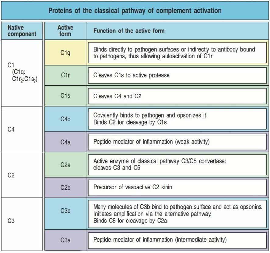
Like the MBL–MASP complex, the C1 complex is made up of a large subunit, called C1q, which acts as the pathogen sensor, and two serine proteases, called C1r and C1s (Figure 11.4). C1q is a hexamer of trimers, formed by monomers that contain an amino-terminal globular domain and a carboxy-terminal collagen-like domain. The trimers assemble through interactions between the collagen-like domains, so that the globular domains join to form a globular head. The complete C1q molecule has six globular heads joined by the collagen-like tails, which allow it to recognise pathogens or antibodies bound to their surface, especially IgG and IgM. C1r and C1s are closely related to MASP-2, while MASP-1 is related to them to a lesser extent. C1r and C1s interact non-covalently to form C1r:C1s pairs. The pairs attach to the arms of C1q, with part of these complexes lying outside C1q.
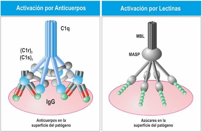
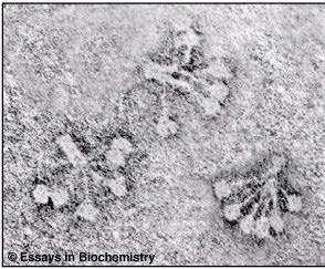
(Reproduced from Regueiro J.R., López C., González S. & Martínez E. (2011) Inmunología. Biología y Patología del Sistema Inmune. (4ª Ed.) Editorial Médica Panamericana, Madrid. Reproduced
from Murphy K et al. (2012) Janeway’s Immunobiology (8th Ed.) Garland Science, New York.)
The binding of two or more C1q heads to their ligand produces a conformational change in the C1r:C1s complex, which activates the autocatalytic activity of C1r. C1r then cleaves its associated C1s protein to generate an active serine protease. C1s acts on the next two components of the classical pathway, C4 and C2. C1s cleaves C4 to produce C4b, which binds covalently to the surface of the pathogen, as in the lectin pathway. A molecule of C2 binds to C4b and is cleaved by C1s, releasing the serine protease C2b. This process leads to the formation of C4b2b, which is the C3 convertase of the classical and lectin pathways (Figure 11.5 and Figure 11.6).
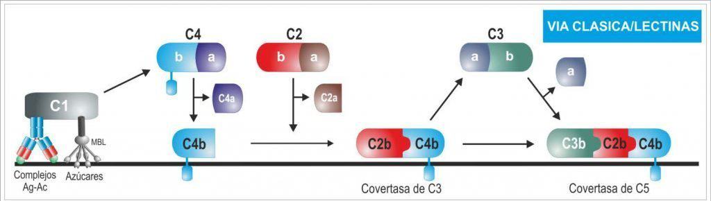
(Reproduced from Regueiro J.R., López C., González S. & Martínez E. (2011) Inmunología. Biología y Patología del Sistema Inmune. (4ª Ed.) Editorial Médica Panamericana, Madrid.)
(Reproduced from Murphy K et al. (2012) Janeway’s Immunobiology (8th Ed.) Garland Science, New York.)