Here is the syllabus explained in the video above. If you have any questions, post them in the comments section at the end of the page.
The cells that take part in the innate immune response carry out different functions to eliminate pathogens:
- Phagocytosis and lysis (Figure 2.13, left): phagocytes can recognise foreign substances by means of receptors on their membrane surface. Phagocytes engulf pathogens, forming a phagocytic vesicle that fuses with a primary lysosome, whose enzymes digest the pathogen. In this way a phagosome or secondary lysosome is formed. Phagocytosis and lysis is the main function of macrophages, which are responsible for delayed hypersensitivity. Neutrophils also phagocytose in response to signals such as complement and antibodies. Eosinophils can acquire phagocytic capacity in certain circumstances, although their main function is the response to parasites.
- Exocytosis and damage (Figure 2.13, centre): eosinophils can recognise parasites, triggering a signalling cascade that induces the release of substances that are cytotoxic and neurotoxic to the parasite.

The innate destruction of small pathogens (bacteria) or large ones (parasites) involves different cells. Inflammation helps to recruit other cells and molecules. (Reproduced from Regueiro J.R., López C., González S. & Martínez E. (2011) Inmunología. Biología y Patología del Sistema Inmune. (4th Ed.) Editorial Médica Panamericana, Madrid.)
- Exocytosis and inflammation (Figure 2.13, right): mast cells and basophils recognise antigens by means of an immunoglobulin E anchored on their membrane surface. Recognition leads to the secretion of a collection of preformed inflammatory molecules stored in the granules, which produce effects including vasodilatation, extravasation of cells from the bloodstream into the infected tissue, chemotaxis and oedema (swelling).
- Missing-self surveillance: NK cells detect the loss of cellular expression of certain proteins, usually MHC antigens, and induce cell death by apoptosis. Loss of MHC expression occurs in certain tumour processes and viral infections.
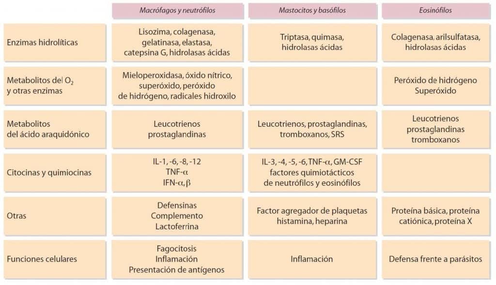
The table shows some substances secreted by macrophages and granulocytes, and their cellular functions. (Reproduced from Regueiro J.R., López C., González S. & Martínez E. (2011) Inmunología. Biología y Patología del Sistema Inmune. (4th Ed.) Editorial Médica Panamericana, Madrid.)
Receptors for antigen recognition
To carry out their functions, immune system cells have a wide variety of receptors on their cell surface. The main receptors are classified below according to their degree of specialisation (Table 2.4):
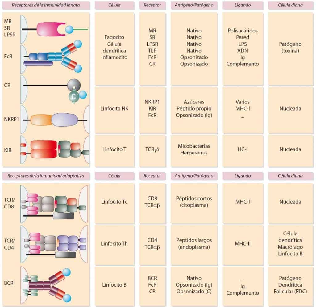
(Reproduced from Regueiro J.R., López C., González S. & Martínez E. (2011) Inmunología. Biología y Patología del Sistema Inmune. (4th Ed.) Editorial Médica Panamericana, Madrid.)
- Receptors for recognition of the molecular patterns of pathogens (PRRs): these are receptors that are not specific for antigen but are specific for a particular chemical structure. They are found on cells of innate immunity, phagocytes, dendritic cells and inflammatory cells. This group includes mannose receptors (MR), bacterial lipopolysaccharide receptors (LPSR or CD14), scavenger receptors (SR), Toll-like receptors (TLRs) and cytoplasmic receptors (NOD or RNA helicases).
- Receptors for adaptor molecules (opsonins): they are found on cells of innate immunity such as phagocytes, dendritic cells, inflammatory cells and NK cells; and on cells of adaptive immunity, such as B cells. The receptor does not recognise the pathogen, but molecules that act as a bridge between the two, called opsonins. Depending on the adaptor molecule recognised, there are two types of receptor: complement receptor (CR), which recognises complement system proteins; and receptor for the constant portion of immunoglobulins (FcR), which recognises the Fc portion of immunoglobulins.
- Two-receptor system: found on NK cells and involved in missing-self surveillance. NK cell activity is based on the balance between the signals emitted by a receptor that activates cell lysis and an inhibitory receptor. There are two important types: immunoglobulin superfamily receptors (KIR, ILT) and lectin-type receptors (CD94).
- Antigen-specific receptors: these correspond to the receptors of T and B cells. They recognise the pathogen in its native state (B cell) or as small peptides within MHC antigens (T cell). T cells are characterised by having a T cell receptor (TcR), of which there is an αβ form (the majority) and a γδ form (the minority). T cells, in turn, can be of two types: helper (Th), which recognise MHC class II molecules and have a CD4 coreceptor, or cytotoxic (Tc), which recognise MHC class I molecules and have a CD8 coreceptor. B cells are characterised by having a B cell receptor (BcR), which includes a membrane immunoglobulin.

(Reproduced from Regueiro J.R., López C., González S. & Martínez E. (2011) Inmunología. Biología y Patología del Sistema Inmune. (4th Ed.) Editorial Médica Panamericana, Madrid.)