Here is the outline explained in the video above. If you have any questions, post them in the comments section at the end of the page.
Types of receptors and ligands
There are essentially two types of phagocytic receptors (Figure 4.4):
- Endocytic receptors, whose function is to mediate phagocytosis.
- Signalling receptors, which induce inflammatory responses against the pathogen.
Recognition of antigens by endocytic receptors, the first large group of phagocytic receptors, leads to internalisation and fusion with lysosomes, where the pathogens are destroyed (Figure 4.4). In addition, in dendritic cells and the monocyte-macrophage system the antigen can also be presented through HLA class II proteins (Figure 4.5), as these are antigen-presenting cells (APCs).
Among the endocytic receptors, the following stand out:
Pattern recognition receptors:
- MR recognises mannose, a carbohydrate that is very common in bacterial and viral glycoproteins and glycolipids.
- LPSR (CD14), together with a TLR, recognises the lipopolysaccharides of Gram-negative bacteria.
- SR recognises debris from Gram-positive bacteria, Gram-negative bacteria or apoptotic cells.
- MBL (mannose-binding lectin) binds to mannose and fucose on bacterial walls and initiates the lectin pathway of complement activation.
- Receptors for adaptor molecules (opsonins): pattern recognition receptors are useless when the pathogen lacks polysaccharides in its capsule. For this reason, such pathogens can be phagocytosed only if they are opsonised by adaptor molecules (immunoglobulins and complement fragments). Phagocytes can recognise opsonised pathogens by means of the following opsonins:
- Immunoglobulin G, recognised by the FcγRI, RII and RIII receptors of phagocytes.
- Complement fragment C3b: recognised by the CR1, CR3 and CR4 receptors of phagocytic cells.
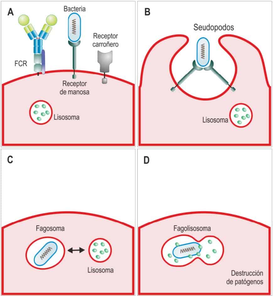
Recognition of microorganisms as foreign by phagocyte receptors leads to their internalisation in phagosomes, which fuse with lysosomes to form phagolysosomes, inside which the microorganisms are destroyed. (Reproduced from Regueiro J.R., López C., González S. & Martínez E. (2011) Inmunología. Biología y Patología del Sistema Inmune. (4th
Ed.) Editorial Médica Panamericana, Madrid.)
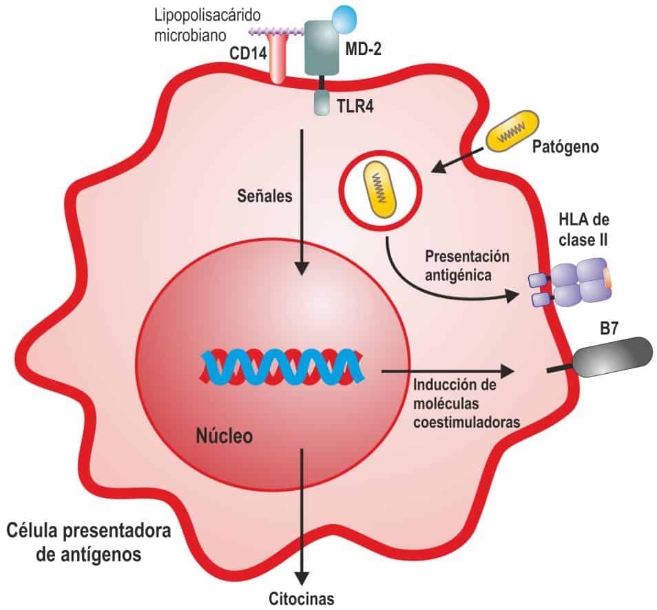
Macrophages take part in inflammation and in the activation of T cells. Recognition of pathogens by signalling receptors (Toll-like receptors, TLRs) transduces a series of signals into the cell nucleus, leading to the synthesis and secretion of cytokines that take part in initiating the inflammatory response and the adaptive immune response. In addition, phagocytosed pathogens are processed and presented as peptides to T cells by MHC or HLA molecules. To be activated, T cells must recognise the foreign peptide and receive a second signal from a series of costimulatory molecules that are expressed on the surface of phagocytes when signalling receptors recognise microorganisms. (Reproduced from Regueiro J.R., López C., González S. & Martínez E. (2011) Inmunología.
Biología y Patología del Sistema Inmune. (4th Ed.) Editorial Médica Panamericana, Madrid.)
Signalling receptors, which form the second large group of phagocytic receptors, are distributed on the surface, in endosomes, or even free in the cytoplasm of the cell. They recognise pathogen-associated molecular patterns (PAMPs), and there are several families of this type of receptor, one of the most important being the Toll-like receptors (TLRs).
Toll-like receptors (TLRs)
Toll-like receptors have an extracellular domain containing many leucine-rich repeats (LRRs), a transmembrane domain and an intracellular domain, or TIR domain (Figure 4.6). The TIR domain interacts with other members of the TLR signal transduction pathway, such as MyD88 (Figure 4.7).
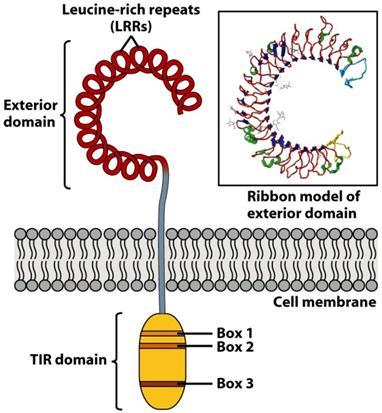
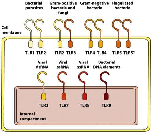
(Reproduced from Kindt T.J., Goldsby R.A., Osborne B.A. & Palacios R. (2007) Inmunología de Kuby (6th Ed.) McGraw-Hill Interamericana, Madrid.)
Ten TLRs are known in humans, each able to recognise a different set of pathogen molecular patterns (PAMPs) that do not normally exist in vertebrates. Because there are only 10 different proteins, their specificity is limited, but they can recognise molecules from very different pathogens.
Some TLRs are expressed on the cell surface (those that recognise proteins, peptidoglycans, lipoproteins or LPS), while others are expressed in the endosomal membrane (Figure 4.7), where they detect fragments of pathogens that have previously been endocytosed.
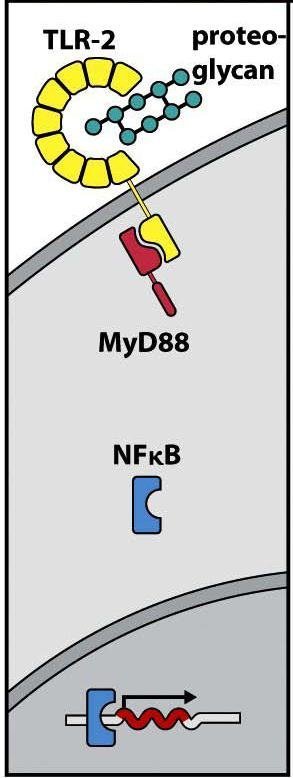
Recognition of proteoglycans by TLRs 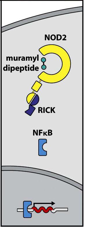
Recognition of proteoglycans by NOD
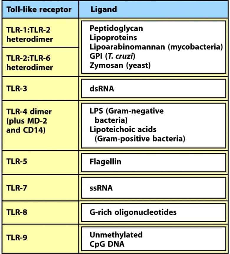
(Reproduced from Murphy K et al. (2008) Janeway’s Immunobiology (7th Ed.) Garland Science, New York.)
Endosomal TLRs mainly recognise different forms of nucleic acids from microorganisms. They can act as heterodimers, homodimers, monomers or, in the case of TLR-4, through association with the proteins CD14 and MD-2 to recognise LPS bound to an LPS-binding protein (acute phase of an infection) (Figure 4.8).
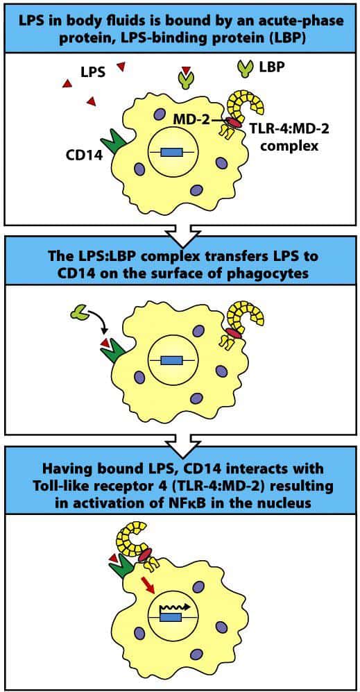
(Reproduced from Murphy K et al. (2008) Janeway’s Immunobiology (7th Ed.) Garland Science, New York.)
Another family of receptors similar to TLRs but with a cytosolic distribution are the NOD (nucleotide-binding oligomerisation domain) receptors, which bind bacterial products free in the cytoplasm (Figure 4.7). Signalling induced by TLRs and NOD has some distinct mediators and some shared ones. In both cases, however, the nuclear transcription factor NF-κB is activated, translocates to the nucleus and induces the synthesis of cytokines and other proteins of the inflammatory process (Figure 4.9).
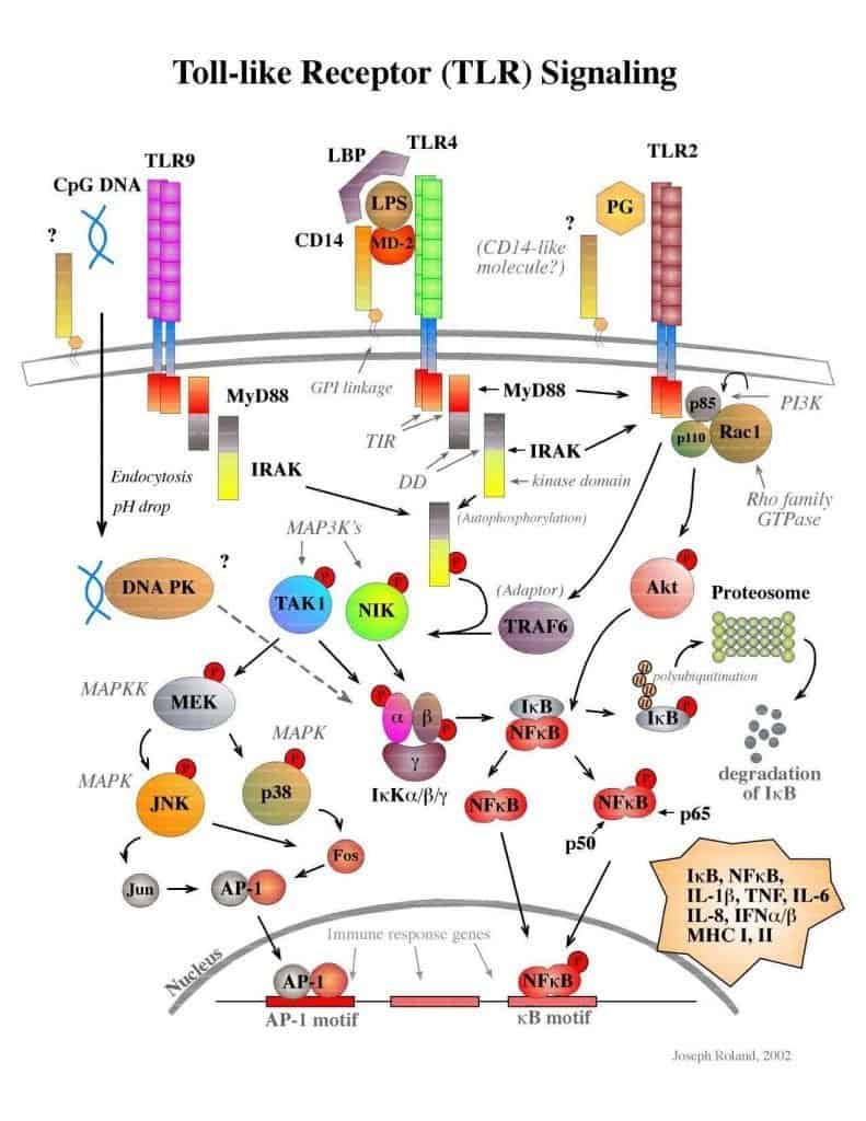
(Courtesy of Joseph Roland.)