What is the pericardium?
The pericardium is a fibroserous sac that surrounds the heart and the roots of the great vessels. In the pericardium we can distinguish two parts:
- Serous pericardium
- Fibrous pericardium

Remember that this lesson is part of the free courses from Paradigmia. Access the full course below 👇👇 HUMAN ANATOMY COURSE
Serous pericardium
This is a very thin, transparent double membrane made up of two layers:
- Visceral layer or epicardium
- Parietal layer
The visceral layer
This layer directly lines the heart and the roots of the vessels that leave or enter it. It is also in direct contact with the myocardium.
In some areas between the visceral layer or epicardium and the myocardium there may be adipose tissue, especially at the atrioventricular or coronary groove and in the interventricular grooves. The coronary arteries and their most important branches run along these grooves.
You can review the coronary vessels in the next lesson 👇
The parietal layer
This layer lies more superficially than the visceral one, and a small space is left between the two layers, the pericardial cavity
This cavity contains a thin film of pericardial fluid. The function of this fluid is to keep the surfaces of the serous membranes moist so as to make it easier for the layers to glide over each other during the movements of the heart.
When the serous pericardium becomes inflamed, pericarditis occurs. The causes of pericarditis are varied:
- Viral infections
- Rheumatic fevers
- Tuberculosis
- Metabolic disorders
- Etc...
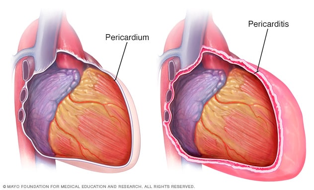
If there is pericarditis, a pericardial effusion may develop. A pericardial effusion is an increase of fluid in the pericardial cavity. This causes a condition with retrosternal pain, and if the effusion is large it can hinder the movements of the heart and the filling of the atria by compression (cardiac tamponade).

Blood can also accumulate (haemopericardium) because of the rupture of a vessel or because of wounds that affect the thoracic cavity.
Reflections of the pericardium
The parietal and visceral layers of the serous pericardium continue into each other in certain areas known as zones of reflection (of the visceral layer onto the parietal layer).
Seen from the front, the reflection of these two layers is easy to see. It occurs at the level of the anterior surface of the aorta and the pulmonary artery and of the superior vena cava (the initial segment of these vessels is intrapericardial).
Transverse sinus or Theile’s sinus
Behind the intrapericardial part of the aorta and the pulmonary trunk there is a tunnel of visceral pericardium known as the transverse sinus or Theile’s sinus. This tunnel is bounded by:
- In front, by the aorta and the pulmonary artery.
- Superiorly, by the right branch of the pulmonary artery.
- Inferiorly and posteriorly, by the atria.

The transverse sinus has two entrance orifices:
- The right orifice: it lies between the superior vena cava laterally, the origin of the aorta medially and the right atrial appendage below.
- The left orifice: it is bounded by the origin of the pulmonary artery medially and by the left atrium and left atrial appendage laterally and below.
Oblique sinus or Haller’s sinus
Posteriorly, the visceral pericardium lines the atria (the base of the heart) and reflects to continue with the parietal layer at the level of the entry of the pulmonary veins into the left atrium and of the inferior vena cava into the right atrium.
Taken as a whole, this zone of reflection has a U shape that opens downwards, and a recess or cul-de-sac is formed between the parietal and visceral layers, called the oblique sinus or Haller’s sinus.


Fibrous pericardium
It is a fibrous, resistant sac that adheres superficially to the parietal layer of the serous pericardium. Superiorly, the fibrous pericardium extends slightly beyond the serous pericardium and ends by fusing with the adventitia (outer connective tissue layer) of the vessels entering and leaving the heart.
The fibrous pericardium is attached to nearby structures by tracts of dense connective tissue known as pericardial ligaments:
- Sternopericardial ligaments: they stretch from the pericardium to the sternum; two are described, one superior and one inferior.
- Vertebropericardial ligament: it runs from the pericardium to the fourth to sixth thoracic vertebra.
- Phrenopericardial ligament: it fixes the pericardium to the central tendon of the diaphragm.