In this lesson of the FREE HUMAN ANATOMY COURSE we are going to study the valves of the heart.
Remember that this lesson is part of the free courses from Paradigmia. Access the full course below 👇👇 HUMAN ANATOMY COURSE
What are the valves of the heart?
The heart is a vital organ made up of four chambers:
- Two atria (upper chambers)
- Two ventricles (lower chambers)

Functions of the heart valves
While the heart is working, blood passes through valves before leaving each chamber for another within the heart. The valves prevent blood from flowing backwards.
In fact, the valves are flaps (leaflets) that act as one-way blood inlets on one side of the ventricle and as one-way blood outlets on the other side of the ventricle.
In the following image you can see a heart at work, and, as you will see, the heart valves open and close in synchrony with the cardiac cycle.

Normal valves have three flaps (leaflets), except the mitral valve, which has only two. The four valves of the heart are the following:
- Tricuspid valve
- Pulmonary valve
- Mitral valve
- Aortic valve
We will now look in depth at each of these valves:
Tricuspid valve
The tricuspid valve, or right atrioventricular valve, is on the right dorsal side of the mammalian heart, at the upper part of the right ventricle.
The function of the valve is to prevent backflow (regurgitation) of blood from the right ventricle into the right atrium during contraction of the right ventricle: systole.
Pulmonary valve
The pulmonary valve is the semilunar valve of the heart that lies between the right ventricle and the pulmonary artery, and it has three cusps. Like the aortic valve, the pulmonary valve opens in ventricular systole, when the pressure in the right ventricle rises above the pressure in the pulmonary artery.
At the end of ventricular systole, when the pressure in the right ventricle falls rapidly, the pressure in the pulmonary artery closes the pulmonary valve.
Mitral valve
The mitral valve (also known as the bicuspid valve or left atrioventricular valve) is a valve with two flaps that lies between the left atrium and the left ventricle in the heart.
The mitral valve and the tricuspid valve are known collectively as the atrioventricular valves because they lie between the atria and the ventricles of the heart.
Under normal conditions, blood flows through the open mitral valve during diastole with contraction of the left atrium, and the mitral valve closes during systole with contraction of the left ventricle.
The valve opens and closes because of pressure differences: it opens when the pressure in the left atrium is higher than in the ventricle, and closes when the pressure in the left ventricle is higher than in the atrium.
Under abnormal conditions, blood can flow backwards through the valve (mitral regurgitation) or the mitral valve can become narrowed (mitral stenosis).
Let us look at these two conditions in the following images:
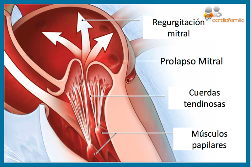

Rheumatic heart disease often affects the mitral valve; the valve can also prolapse with age and be affected by infective endocarditis. The mitral valve is named after a bishop’s mitre, which its flaps resemble.
Aortic valve
This valve is located between the left ventricle and the aorta.
It is one of the two semilunar valves of the heart; the other is the pulmonary valve.
The aortic valve normally has three cusps or leaflets, although in 1-2% of the population it is found to have two leaflets congenitally. Let us look at its anatomy at work in the following image:

The aortic valve is the last structure of the heart that blood travels through before the flow passes into the systemic circulation.
Remember that this lesson is part of the free courses from Paradigmia. Access the full course below 👇👇 HUMAN ANATOMY COURSE