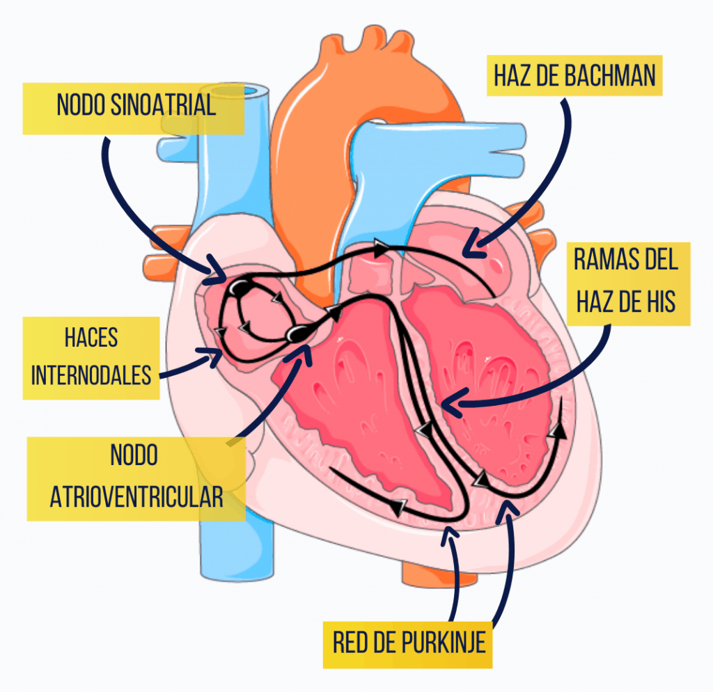
This is a series of highly differentiated fibres that are able to generate electrical impulses autonomously (the system is said to be self-exciting) and to transmit these impulses to the rest of the myocardium, so that the muscle fibres contract progressively and in synchrony. In this way it regulates the heart rate and orders the contraction of the myocardium.
In this conduction system we can consider a number of parts:
Sinoatrial node
Under normal conditions this node initiates the cardiac impulse (which is why it is called the pacemaker of the heart) and determines the heart’s rate of contraction. It is located in the right atrium, lateral to the opening of the superior vena cava, and generates impulses at a rate of 60 or 70 impulses per minute, but this rate can be modified by nervous influence (autonomic nervous system) or hormonal influence (catecholamines). If the sinoatrial node fails, an artificial pacemaker has to be implanted, which produces electrical impulses that initiate the ventricular contractions at a previously established rate. The pacemaker is usually implanted by right heart catheterisation (generally through the brachial vein); the catheter carries an electrode that is inserted into the trabeculae carneae of the right ventricle.
Internodal tracts
There are three of them, anterior, middle and posterior, and they send the impulse from the sinoatrial node to the atrioventricular node.
Another bundle is described that, starting from the sinoatrial node, crosses the interatrial septum and reaches the left atrium: this is the bundle of Bachmann.
Atrioventricular node
It is located in the right atrium, in the posteroinferior part of the interatrial septum, close to the opening of the coronary sinus. It also has the capacity to generate impulses, but at a lower rate than the sinoatrial node. As we have seen, it is connected to the sinoatrial node through the internodal tracts, and under normal conditions the activity of the atrioventricular node is masked by that of the sinoatrial node; but if the latter fails, the atrioventricular node can work autonomously, although generating impulses at a lower rate. Under normal conditions, the propagation of the impulse is slightly delayed in the atrioventricular node, which allows atrial contraction to take place before ventricular contraction.
Atrioventricular bundle or bundle of His
This bundle starts from the atrioventricular node and passes through the right fibrous trigone to reach the interventricular septum; shortly afterwards it divides into two branches, right and left. The right branch of the bundle of His runs along the septomarginal trabecula or arciform band, and the left branch crosses the interventricular septum to reach the left ventricle.
The two branches of the bundle of His branch out through the ventricles, forming the so-called Purkinje network, which carries the electrical stimulus to the ventricular muscle fibres so that they contract.
The transmission of electrical impulses through the heart can be amplified and recorded by means of the electrocardiogram.
Many heart problems cause conduction disturbances of the cardiac electrical stimulus, and these can be detected by means of the electrocardiogram.