In this lesson we will look at what the larynx is, the cartilages of the larynx, their joints and its ligaments.
Remember that this lesson is part of the free courses from Paradigmia. Access the full course here ▶️ HUMAN ANATOMY COURSE ◀️
- What is the larynx?
- Cartilages of the larynx
- Cartilages of the larynx: thyroid
- Cartilages of the larynx: cricoid
- Cartilages of the larynx: epiglottic
- Cartilages of the larynx: arytenoid
- Cartilages of the larynx: corniculate
- Cartilages of the larynx: cuneiform (or Morgagni’s)
- Joints of the larynx
- Cricothyroid joint
- Cricoarytenoid joint
- Ligaments of the larynx
- Recommended reading on the cartilages of the larynx
What is the larynx?
The larynx is the first individualised portion of the airway. It is a mechanism that separates the digestive and respiratory tracts, and it is highly specialised for phonation.
The larynx is an unpaired, median structure in the neck that is easily accessible to surface examination; it lies between the 3rd and 6th cervical vertebrae in men, and higher in women and children.
In men the thyroid cartilage is larger and more prominent than in women as a result of greater post-pubertal development, which gives them a deeper voice.
The larynx is held in place by its continuity with the hyoid bone above and with the trachea below; it is also quite mobile in the vertical direction thanks to the suprahyoid and infrahyoid muscles.
Cartilages of the larynx
Within the anatomy of the larynx we find the following components: a cartilaginous skeleton, joints and ligaments between the cartilages, muscles, and an inner mucosa with a series of characteristic features.
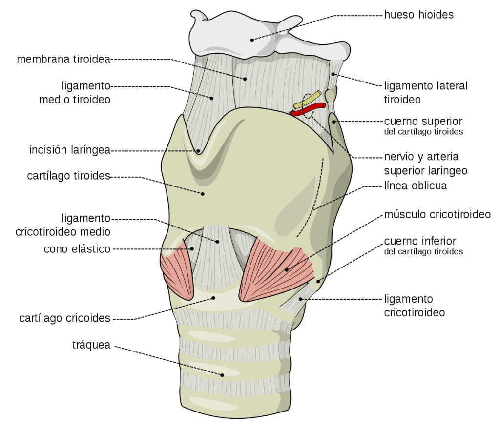
Cartilages of the larynx: thyroid
This is the largest and protects the rest. It consists of two quadrilateral laminae that join anteriorly to form the laryngeal prominence, or Adam’s apple, except in their upper part, where a gap, the superior thyroid notch, is left.
The posterior border of each lamina is thick and vertical, and extends into projections, or superior and inferior horns, of which the superior is much more pronounced; on the inner surface of the inferior horns there are articular surfaces for joining the cricoid.
Cartilages of the larynx: cricoid
The rest of the cartilage is known as the arch and has an anterior median tubercle and two lateral articular surfaces for articulation with the thyroid.
It lies between the larynx and the trachea. It is ring-shaped, with a thick posterior part called the cricoid lamina, or plate, on whose upper surface there are two elliptical articular surfaces (facing upwards, forwards and outwards) for articulation with the arytenoid cartilages.
Cartilages of the larynx: epiglottic
It is unpaired and leaf-shaped, with a narrowed lower part that attaches to the inner angle of the thyroid cartilage. On the posterior surface there are numerous depressions produced by glands of the laryngeal mucosa.
Cartilages of the larynx: arytenoid
These are two triangular pyramids that articulate by their base with the lamina of the cricoid. They have an anterolateral surface with the triangular fovea and, below it, the oval fovea, separated by the arcuate crest.
The base has an elliptical articular surface for articulation with the cricoid and has two projections: one anteromedial, or vocal process (which gives
attachment to the vocal ligament), and one posterolateral, or muscular process.
Cartilages of the larynx: corniculate
Also called the cartilages of Santorini, these are two small cartilages that articulate with the apex of the arytenoids.
Cartilages of the larynx: cuneiform (or Morgagni’s)
They lie in front of and lateral to the corniculate cartilages, in the aryepiglottic folds. They are inconstant.
Joints of the larynx
Cricothyroid joint
It occurs between the inferior horns of the thyroid cartilage and the cricoid lamina. It is a plane (gliding) joint in which the thyroid tilts on the cricoid in an anteroposterior direction (which tenses the vocal cords).
Cricoarytenoid joint
It lies between the upper surface of the cricoid lamina and the base of each arytenoid. The cricoid articular surface slopes upwards, outwards and forwards, so that at this level the arytenoid undergoes two types of sliding movement, one outwards and downwards and the other inwards and upwards, which alters the rima glottidis.
In addition, there is a rotation about a vertical axis, so that the vocal processes and vocal cords move towards and away from each other.
Ligaments of the larynx
The space between the thyroid cartilage and the hyoid bone is closed by the thyrohyoid membrane, which extends from the upper border of the thyroid cartilage to the lower border of the body and greater horns of the hyoid.
The cricothyroid membrane lies anteriorly and runs from the lower border of the thyroid cartilage to the upper border of the cricoid ring; it is thick and elastic, and it is the site of choice for emergency access to the airway in laryngeal obstruction.
The cricotracheal membrane is a lamina of connective tissue that joins the cricoid to the first tracheal ring.
The thyroepiglottic ligament is a fibrous band that joins the apex of the epiglottis to the inner surface of the angle of the thyroid cartilage.
The intrinsic ligaments of the larynx include:
- The vocal ligaments (true vocal cords), or inferior thyroarytenoid ligaments, run from the vocal process of the arytenoid to the re-entrant angle of the thyroid cartilage; they continue downwards as a membrane that is fixed to the upper border of the cricoid, the conus elasticus.
- The hyoepiglottic ligament is a median fibrous lamina that joins the upper surface of the epiglottis to the posterosuperior border of the hyoid bone (being elastic, it helps to raise the epiglottis during inspiration). We have already studied the median and lateral glossoepiglottic folds.
- The vestibular ligament (false vocal cord), or superior thyroarytenoid ligament, runs from the re-entrant angle of the thyroid cartilage to the oval fovea of the arytenoid and continues upwards as a fibrous lamina known as the quadrangular (or elastic) membrane, which extends until it fuses with the aryepiglottic fold.
Remember that this lesson is part of the free courses from Paradigmia. Access the full course here ▶️ HUMAN ANATOMY COURSE ◀️