The parasympathetic innervation of the thoracic viscera comes from the vagus nerves.
These nerves reach the thorax from the neck, accompanying the common carotid arteries and the internal jugular veins (neurovascular bundle of the neck). In the thorax their course differs on the right and left sides.
Right vagus
It crosses in front of the right subclavian artery, and at this level gives off the right inferior laryngeal (recurrent) nerve , which loops under and behind the subclavian artery and ascends in the neck lateral to the oesophagus. After crossing the subclavian artery, the right vagus nerve descends through the superior mediastinum and runs backwards, crosses the trachea obliquely and passes behind the right root of the lung. Finally, in the posterior mediastinum it reaches the oesophagus and lies behind and to the right of it; at this level it divides into numerous fascicles that help to form the oesophageal plexus.

Left vagus
It enters the thorax between the left common carotid artery and the left subclavian artery, descends through the superior mediastinum, reaches the arch of the aorta and crosses it on its left lateral surface. In the lower part of the arch it gives off the left inferior laryngeal (recurrent) nerve, lateral to the ligamentum arteriosum. The left inferior laryngeal nerve loops under the arch of the aorta and then ascends through the tracheo-oesophageal groove towards the neck.
After crossing the arch of the aorta, the left vagus runs downwards and backwards and passes behind the left root of the lung, reaching the posterior mediastinum to arrive at the oesophagus and lie in front of and to the left of it. Like the right vagus, it divides into numerous fascicles that help to form the oesophageal plexus.
In the neck, the vagus nerves give off the superior and middle cardiac vagal nerves and in the thorax they give the inferior cardiac vagal nerve , which, together with the sympathetic cardiac nerves, reach the cardiac plexus.
- The cardiac vagal nerves carry sensory fibres and preganglionic motor fibres. The latter synapse in small parasympathetic ganglia located in the cardiac plexuses, and postganglionic fibres leave these ganglia for the heart.
- The efferent (motor) fibres of the parasympathetic system reduce the heart rate (bradycardia) by slowing conduction in the conduction system of the heart. Parasympathetic stimulation, like sympathetic stimulation, occurs reflexly.
- The afferent (sensory) fibres mainly carry baroreceptor information from the arch of the aorta (these impulses "inform" the central nervous system about blood pressure).
The vagus nerves also give branches to the pulmonary plexus, located around the roots of the lungs and the tracheal bifurcation.
- The efferent (motor) fibres of this plexus are preganglionic. They reach the tracheobronchial tree and synapse in small parasympathetic ganglia in its vicinity or within the wall itself; postganglionic fibres leave these for the smooth muscle, glands and blood vessels. Parasympathetic stimulation causes contraction of the tracheal and bronchial smooth muscle (bronchoconstriction) and increased glandular secretion.
- The afferent (sensory) fibres carry information about the degree of alveolar distension and about stimuli that irritate the tracheal or bronchial mucosa; these stimuli act as the afferent limb of the cough reflex. These sensory fibres run from the trachea and bronchi to the pulmonary plexus and then join the vagus nerves.
The parasympathetic innervation of the oesophagus comes from the vagus nerves. These nerves carry preganglionic fibres that synapse in small parasympathetic ganglia located in the muscular layer (myenteric plexus) and in the submucosa (submucosal plexus) of the oesophagus; short postganglionic fibres leave the ganglia for the musculature and glands of the oesophagus. The parasympathetic system stimulates contraction of the oesophageal muscle and glandular secretion.
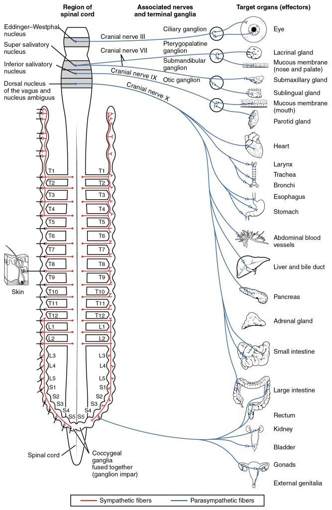
Source: Anatomy and Physiology, Open Stax CNX