The lungs are essential organs for human function; gas exchange, also known as haematosis, takes place in them. In this process, venous blood (rich in carbon dioxide) arrives at the lungs and is transformed into arterial blood (rich in oxygen); this process is known as haematosis.
They are located in the thoracic cavity, enveloped by a double serous membrane, the pleurae. Inferiorly they are separated from the abdominal cavity by the diaphragm, and the two lungs are separated from each other by viscera located in the thoracic mediastinum. The right lung is bulkier than the left.
External features and anatomical relations
In each lung we can distinguish three surfaces:
- Costal, in contact with the wall of the thorax.
- Medial or mediastinal, facing the thoracic mediastinum.
- Inferior or diaphragmatic, facing the diaphragm.
Separating these surfaces, three borders are described:
- Anterior border, separating the costal surface from the mediastinal surface in front. In the left lung, in the lower part of this border there is a notch produced by the heart: the cardiac notch.
- Posterior border, separating the costal and mediastinal surfaces behind.
- Inferior border, separating the diaphragmatic surface from the other two surfaces.
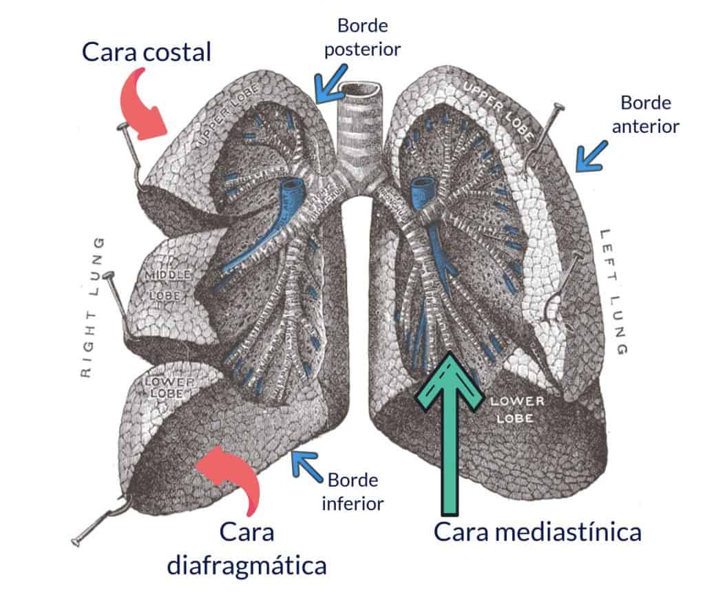
The uppermost part of the lung, which ends in a rounded point, is called the apex of the lung; it projects partly above the superior thoracic aperture and reaches the supraclavicular region, so it can be injured in wounds at the base of the neck.
Fissures and lobes
On the surface of the right lung two broad fissures can be seen, one called the horizontal or minor fissure and the other the oblique or major fissure. This allows us to distinguish in the right lung three parts lying between the fissures: the superior, middle and inferior lobes of the lung.
The left lung has only one fissure, the oblique fissure, so it has only two lobes, superior and inferior; it has no middle lobe.

Occasionally accessory fissures can be seen, which correspond to lines of demarcation of the bronchopulmonary segments (we will analyse this concept later).
There is a fissure that appears fairly often in the right lung, produced by the azygos vein. In these cases the vein ascends higher than normal and sinks into the lung parenchyma, marking a deep groove at the apex of the lung called the azygos fissure. This gives rise to an accessory lobe, known as the azygos lobe, which occurs in 1% of individuals. The azygos fissure can be seen on a radiograph as a linear mark that separates part of the apex of the lung from the rest of the lung.

Costal surface
It is convex and is the broadest surface in both lungs. It is related, through the pleura, to the ribs and intercostal spaces.
On the right side, both fissures (horizontal and oblique) are seen on this surface, so the three lobes (superior, middle and inferior) can also be distinguished.
In the left lung there is only one fissure (oblique), and we can see two lobes (superior and inferior) on this surface. The part of the superior lobe that lies below the cardiac notch is known as the lingula of the left lung.
Medial or mediastinal surface
It is slightly concave, and it contains the hilum of the lung, the area of the lung through which the elements of the pulmonary pedicle enter or leave. The main bronchus, the pulmonary artery, the bronchial artery or arteries and nerves enter through the hilum. The pulmonary veins and the lymphatic vessels of the lung leave through the hilum.
On this surface of the right lung, both fissures (horizontal and oblique) can also be seen, converging at the hilum. We can also see the three lobes: the superior lobe located above the hilum, the middle lobe in front of the hilum, and the inferior lobe behind and below the hilum.
In the left lung we see only the oblique fissure, interrupted at the hilum; as we know, this fissure separates the superior and inferior lobes.
In relation to the position of the hilum, we distinguish three portions on this surface: the prehilar portion, in front of the hilum; the retrohilar portion, behind the hilum; and the suprahilar portion, above the hilum. We are going to analyse the relations of each portion.
In the right lung:
- The prehilar portion is related to the heart, which produces a depression on this surface known as the cardiac impression.
- The retrohilar portion is related to the oesophagus and to the azygos vein; both structures can mark two longitudinal grooves behind the hilum of the lung.
- The suprahilar portion is related to the arch of the azygos vein, which forms a groove that curves over the top of the hilum. Above this groove, the suprahilar portion is related, from front to back, to the right brachiocephalic vein, the trachea and the oesophagus; all three can leave vertical grooves on the mediastinal surface of the right lung.
In the left lung, the mediastinal surface is related:
- The prehilar portion is related to the heart, which produces the cardiac impression, more marked than in the right lung. Anteriorly, below the cardiac notch, the lingula of the lung can be seen.
- The retrohilar portion is related to the oesophagus and, further back, to the descending thoracic aorta; both, but above all the aorta, can leave vertical grooves behind the hilum.
- The suprahilar portion is related above all to the arch of the aorta, which leaves a deep groove that curves over the top of the hilum. Above it, three vertical grooves can be seen, from front to back: the groove for the left brachiocephalic vein, the groove for the left subclavian artery, and the groove for the trachea and the oesophagus.
It is important to point out that the grooves and all the anatomical features described above are well defined only in fixed lungs, and they are not always clearly seen.
Diaphragmatic surface
It is concave, and in both lungs the oblique fissure can be seen on this surface: on the right side it separates the middle lobe, which lies anteriorly, from the inferior lobe, which is more posterior; on the left side the oblique fissure separates the superior lobe (more anterior) from the inferior lobe. This surface adapts to the corresponding dome of the diaphragm, but posteriorly the diaphragm descends further than the lung.
Through the diaphragm, the right lung is related mainly to the liver, and the left lung to the stomach and the spleen.
When clinicians talk about the base of the lung, they do not mean the inferior or diaphragmatic surface of the lung, but rather the most posterior and inferior portion of the lung, which is auscultated by placing the stethoscope on the posterior wall of the thorax at the level of the tenth rib.
Apex
It extends slightly beyond the first rib, partly reaching the supraclavicular region. It is important to know these relations because lung cancers very frequently affect the apex.
- Vascular relations: the subclavian artery lies above the apex of the lung and the common carotid artery medial to it.
- Nervous relations: posteriorly, the apex of the lung is related to the cervicothoracic (stellate) sympathetic ganglion; anteriorly to the phrenic and vagus nerves; superiorly to the brachial plexus; and medially to the inferior laryngeal nerves.
- Visceral relations: medially, the apex is related to the trachea and the oesophagus.
Regarding the nervous relations above:
The relation between the apex and the cervicothoracic sympathetic ganglion explains why autonomic symptoms such as tachycardia, constriction of the pupil (miosis), and redness and sweating disturbances on one half of the face (Bernard-Horner syndrome) can appear in some cancers of the apex of the lung; these symptoms are produced by injury or irritation of the cervical sympathetic nervous system.
The relation between the apex and the phrenic and vagus nerves anteriorly is important because some tumours of the apex can injure the phrenic nerve and cause paralysis of the corresponding hemidiaphragm.
The relation between the apex and the brachial plexus superiorly explains why motor and sensory symptoms in the upper limb can appear in tumours of the apex of the lung.
Medially, the apex is related to the inferior laryngeal nerves (recurrent), so voice disturbances (aphonia or dysphonia) can appear in cancers of the apex of the lung.
